Here is another short excerpt from my recently published book, Neoplasms: Principles of Development and Diversity.
"Many more people are alive today as the result of precancer treatment than are alive due to the treatment of cancers.
The successful reduction in deaths from cervical cancer provides a good example of the effectiveness of precancer treatment. Cervical cancer is a type of squamous cell carcinoma that develops at the junction between the ectocervix (the squamous lined epithelium) and the endocervix (the glandular lined epithelium) in the os of the uterine cervix of women. Before the introduction of cervical precancer treatment, cervical carcinoma was one of the leading causes of cancer deaths in women. Today, in many countries that have not deployed precancer treatment, cervical cancer is the leading cause of cancer deaths in women (72, 73). The relatively low number of cervical cancer deaths in the United States is the result of a 70% reduction in age-adjusted mortality after the introduction of Pap smear screening (74–76). No effort aimed at treating invasive cancers has provided an equivalent reduction in the number of cancer deaths as this simple procedure for treating precancers.
Today, we know that almost all cervical cancer is due to infection by one of several carcinogenic strains of human papillomavirus. The strains of human papillomavirus that cause cervical cancer are transmitted during sexual intercourse by men infected with the virus. In the late 1940s (and really up until the early 1980s), the viral etiology of cervical cancer was unknown. We did know that morphologic changes in cervical epithelial characterized the early steps in cervical cancer development. By sampling and examining cervical specimens from women, it was possible to accurately determine whether precancerous changes were present. If precancerous changes were present, a gynecologist could remove a superficial portion of the affected epithelium, and this would, in the vast majority of cases, stop the cancer from ever developing.
Thanks largely to the persistence of Dr. Papanicolaou and his coworkers, a screening test was developed to detect cervical precancers. The Pap smear is obtained by scraping or brushing the junction between the endocervix and the ectocervix and
spreading the detached cells onto a glass slide. The cells are then stained with a histologic reagent (the Papanicolaou stain) that allows the cytologist to visualize subtle alterations in cell morphology. A typical Pap smear contains about 20,000 cells, and every cell must be inspected to rule out dysplasia and other pathologic abnormalities.
A large cytology laboratory can handle hundreds of thousands of Pap smears in a year. In the last half of the 20th century, the Pap smear cytologic evaluation led to at least a 70% drop in the number of deaths from cervical cancer in every country that fully deployed the test.
Morphologic and epidemiologic observations on Pap smears provided clues that led to the identification of several strains of human papillomavirus as the major causes of cervical cancer. [Half of the The 2008 Nobel Prize in Medicine went to Harald zur Hausen, for his work, showing the relationship between human papillomavirus and cervical cancer]. Recently, a vaccine against carcinogenic strains of HPV has been developed. Gardasil and Cervarix are two HPV vaccines that are currently available. If all goes well, most cases of cervical cancer will be prevented by a cancer vaccination. The Pap smear industry will shrink as fewer and fewer women test positive for cervical dysplasia.
The examination of Pap smears is just one of the many activities of a cytology laboratory. Cytologists routinely diagnose cells obtained from virtually any anatomic site and from any body fluid."
- Jules Berman
Friday, November 28, 2008
Thursday, November 27, 2008
Thanks for all the data!
The United States is celebrating Thanksgiving today. Thanksgiving is my favorite holiday, because it allows everyone, regardless of their religion or personal philosophy, to join together as a nation in thanks for the many wonderful things this world provides.
This year is a particularly great Thanksgiving, because we have a newly elected President. Billions of people throughout the world hope that our new leadership will contribute to multinational efforts to reduce global warming, to use resources wisely and fairly, and to reduce violence and disease.
As someone who specializes in the field of biomedical informatics, I am personally grateful for the infrastructure that permits me to contribute to society, just by sitting at my computer and using the publicly available digital resources.
Here is the list of free resources that I am particularly grateful for (many of which accept charitable contributions):
Searchable information
PubMed
Wikipedia
Google
Google Earth
Languages and software
Perl
Ruby
Python
R
ImageMagick
OpenOffice
Datasets
SEER
Entrez
U.S. Census
Taxonomy.dat
OMIM
NASA images
Nomenclatures and ontologies
MESH
ICD
NCI Thesaurus
GO
Standards and protocols
HTML
XML
RDF
HTTP
FTP
JPEG
PNG
Organizations
GNU
OBO
CPAN
There are many many others, but these are all that I can think of at the moment. Please feel free to add your favorite free data resources, for which you are particularly thankful.
Happy Thanksgiving,
-Jules Berman
This year is a particularly great Thanksgiving, because we have a newly elected President. Billions of people throughout the world hope that our new leadership will contribute to multinational efforts to reduce global warming, to use resources wisely and fairly, and to reduce violence and disease.
As someone who specializes in the field of biomedical informatics, I am personally grateful for the infrastructure that permits me to contribute to society, just by sitting at my computer and using the publicly available digital resources.
Here is the list of free resources that I am particularly grateful for (many of which accept charitable contributions):
Searchable information
PubMed
Wikipedia
Google Earth
Languages and software
Perl
Ruby
Python
R
ImageMagick
OpenOffice
Datasets
SEER
Entrez
U.S. Census
Taxonomy.dat
OMIM
NASA images
Nomenclatures and ontologies
MESH
ICD
NCI Thesaurus
GO
Standards and protocols
HTML
XML
RDF
HTTP
FTP
JPEG
PNG
Organizations
GNU
OBO
CPAN
There are many many others, but these are all that I can think of at the moment. Please feel free to add your favorite free data resources, for which you are particularly thankful.
Happy Thanksgiving,
-Jules Berman
Sunday, November 23, 2008
Using SEER Public_use Data: 10
This is my tenth and final blog on the SEER Public-Use Data files.
I have prepared three web sites with much of the information that appeared in this series of 10 blog posts:
Obtaining the SEER data files and basic query subroutines, in Perl
A sample project
More examples
If you want to do creative data mining, you will need to learn a little computer programming.
For Perl and Ruby programmers, methods and scripts for using SEER and other publicly available biomedical databases, are described in detail in my prior books:
Perl Programming for Medicine and Biology
Ruby Programming for Medicine and Biology
An overview of the many uses of biomedical information is available in my book,
Biomedical Informatics
More information on cancer is available in my recently published book, Neoplasms
© 2008 Jules Berman
As specified in the SEER Data Agreement, the citation for the SEER data is as follows:
"Surveillance, Epidemiology, and End Results (SEER) Program (www.seer.cancer.gov) Limited-Use Data (1973-2005), National Cancer Institute, DCCPS, Surveillance Research Program, Cancer Statistics Branch, released April 2008, based on the November 2007 submission."
Using SEER Public-Use Data: 9
I have been writing a series of blogs on SEER, the U.S. National Cancer Institute's Surveillance, Epidemiology and End Results program. SEER is an amazing resource for information on the cancers that occur in the U.S. One of the products of SEER is the Public Use dataset, which contains de-identified records on over 3.5 million cancers that have occurred between 1973 and 2005.
When you have 3.5 million cancer cases to study, you can draw certain types of inferences that could not possibly be made with the data accumulated at a single medical institution.
Today, we'll look at the neoplasms that occur in the related anatomic sites: pleura, peritoneum, retro-peritoneum, and pelvis.
Here are the SEER listings. The left-hand column is the number of occurrences. Nneoplasms with fewer than 20 SEER cases were considered un-informative and were omitted from the lists. The second column is the average age of occurrence. The column on the right is the ICD-O term.
SEER captures malignant neoplasms. In the SEER data set, we saw that the following distribution of cases, by occurrences at anatomic site:
The pleura is the mesothelial-lined cavity of the chest, surrounding and covering the heart and the lungs. The peritoneum is the mesothelial-lined cavity of the abdomen, surrounding and overing all or part of the intestines and other organs of the abdomen (e.g., liver, spleen, pancreas).
The pleura accounts for many more occurrences of malignant tumors than does the peritoneum. Only a few different tumors account for the vast majority of malignant neoplasms arising in the pleura.
The peritoneum, with about half as many cancer occurrences as the pleura, has more types of tumors that occur in significant numbers (20 or more), including the tumors that arise from the surface of the ovaries (e.g. papillary serous cystadenocarcinoma).
The retroperitoneum, is the collection of tissues that lie between the peritoneal lining and the surface wall of the abdomen. The retroperitoneum is often referred to as the retroperitoneal space. This is not the best term, as it calls to mind a body cavity (space), perhaps lined by mesothelium, and this is not the case. The retroperitoneum is mostly fat, connective tissue, and organs. Fully retroperitoneal organs, such as the kidney and attached adrenals, can drop a little bit, along the potential space of its surronding fascia, but that's about the closest thing to a space that the retroperitoneum can offer. Organs of the abdomen that are slapped tightly against the posterior wall of the peritoneum are, technically, retroperitoneal (such as the ascending and descending colon, and the rectum). Organs or parts of organs that dangle in the abdomen (such as the transverse colon), are fully peritoneal.

For the purposes of collecting data on retroperitoneal neoplasms, the tumors that arise from identifiable organs (e.g., kidney, head of pancreas, adrenals, rectum) are assigned to those organs, in the SEER dataset, and NOT to the retroperitoneum. This leaves, for the most part, soft tissue tumors, muscle tumors and nerve tumors arising in the retroperitoneum. There are a great variety of these tumors, even when we restrict our list to those tumors that occur with a frequency of 20 or greater.
The pelvis is a commonly used anatomic term that creates much confusion. It is sometimes described as the bowl-like invagination in the lower abdomen, or it may be described as the structures that support the lower abdomen, or it may be described as the set of bones that create the framework of the bowl-like invagination.
It is very difficult to assign neoplasms to the pelvis, because tumors arising in this area can best be assigned to the bones in which they are found, or to the peritoneum, or to the retroperitoneum. The difficulty of assigning neoplasms to the pelvis becomes apparent when we see that of the approximately 3.5 million cases in the SEER dataset, there are only 470 cases assigned to the pelvis, and of these cases, most seem to arise, more specifically, from the peritoneum (papillary serous cystadenocarcinoma) or the uterine cervix (squamous cell carcinoma), or the intestines (adenocarcinoma).
There is a tautologic remark that pathologists use. "Common tumors occur commonly, and uncommon tumors occur uncommonly." This means that a pathologist should be cautious when assigning a diagnosis that rarely occurs at the location where the tumor has arisen.
To know which tumors commonly occur, at what sites, at what ages, in what ethnic populations, it is very useful to have a large collection of neoplasms from which to study, and to have a good understanding of the frequency of occurrence of the neoplasms that arise at the site. The SEER data set permits such determinations.
In a prior blog, I discussed some of the simple Perl routines used in the SEER-data parsing algorithms, and these are available from my web site.
If you want to do creative data mining, you will need to learn a little computer programming.
For Perl and Ruby programmers, methods and scripts for using SEER and other publicly available biomedical databases, are described in detail in my prior books:
Perl Programming for Medicine and Biology
Ruby Programming for Medicine and Biology
An overview of the many uses of biomedical information is available in my book,
Biomedical Informatics.
More information on cancer is available in my recently published book, Neoplasms.
© 2008 Jules Berman
key words: neoplasms, cancer, neoplasia, precancer, tumor, tumour, tumors, tumours, neoplasm, carcinogenesis, carcinogens, tumor genetics, myelodysplastic syndromes, IEN, pre-cancer, preneoplastic lesions, preneoplasia
As specified in the SEER Data Agreement, the citation for the SEER data is as follows:
"Surveillance, Epidemiology, and End Results (SEER) Program (www.seer.cancer.gov) Limited-Use Data (1973-2005), National Cancer Institute, DCCPS, Surveillance Research Program, Cancer Statistics Branch, released April 2008, based on the November 2007 submission."
The image of the retroperitoneum was taken from a reproduction of a Gray's Anatomy image, provided by Wikipedia. The copyright on Gray's Anatomy has expired, and the image is in the public domain.
As with all of my scripts, lists, web sites, and blog entries, the following disclaimer applies. This material is provided by its creator, Jules J. Berman, "as is", without warranty of any kind, expressed or implied, including but not limited to the warranties of merchantability, fitness for a particular purpose and noninfringement. in no event shall the author or copyright holder be liable for any claim, damages or other liability, whether in an action of contract, tort or otherwise, arising from, out of or in connection with the material or the use or other dealings in the material.
In June, 2014, my book, entitled Rare Diseases and Orphan Drugs: Keys to Understanding and Treating the Common Diseases was published by Elsevier. The book builds the argument that our best chance of curing the common diseases will come from studying and curing the rare diseases.

I urge you to read more about my book. There's a generous preview of the book at the Google Books site. If you like the book, please request your librarian to purchase a copy of this book for your library or reading room.
When you have 3.5 million cancer cases to study, you can draw certain types of inferences that could not possibly be made with the data accumulated at a single medical institution.
Today, we'll look at the neoplasms that occur in the related anatomic sites: pleura, peritoneum, retro-peritoneum, and pelvis.
Here are the SEER listings. The left-hand column is the number of occurrences. Nneoplasms with fewer than 20 SEER cases were considered un-informative and were omitted from the lists. The second column is the average age of occurrence. The column on the right is the ICD-O term.
SEER captures malignant neoplasms. In the SEER data set, we saw that the following distribution of cases, by occurrences at anatomic site:
PLEURA = 6,138 cases
PERITONEUM = 3,067 cases
RETROPERITONEUM = 3,640 cases
PELVIS = 470 cases
The pleura is the mesothelial-lined cavity of the chest, surrounding and covering the heart and the lungs. The peritoneum is the mesothelial-lined cavity of the abdomen, surrounding and overing all or part of the intestines and other organs of the abdomen (e.g., liver, spleen, pancreas).
The pleura accounts for many more occurrences of malignant tumors than does the peritoneum. Only a few different tumors account for the vast majority of malignant neoplasms arising in the pleura.
MALIGNANT NEOPLASMS OF THE PLEURA (MALE AND FEMALE)
TOTAL = 6,138 CASES
Number
 of      Age             Neoplasm name
Cases
--------------------------------------------------------------
0024  068   sarcoma nos
0038  071   ml, large b-cell, diffuse
0110  070   neoplasm, malignant
0236  073   mesothelioma, biphasic type, malignant
0433  069   fibrous mesothelioma, malignant
1079  068   epithelioid mesothelioma, malignant
4027  069   mesothelioma, malignant
--------------------------------------------------------------
The peritoneum, with about half as many cancer occurrences as the pleura, has more types of tumors that occur in significant numbers (20 or more), including the tumors that arise from the surface of the ovaries (e.g. papillary serous cystadenocarcinoma).
NEOPLASMS OF THE PERITONEUM (MALE AND FEMALE)
TOTAL = 3067
Number
 of   Age     Neoplasm name
Cases
--------------------------------------------------------------
022  062   liposarcoma nos
023  062   gastrointestinal stromal sarcoma
031  067   sarcoma nos
032  064   carcinoid tumor, malignant
038  085   endometrioid carcinoma
045  061   mucinous adenocarcinoma
046  066   fibrous histiocytoma, malignant
047  067   mullerian mixed tumor
053  066   neoplasm, malignant
073  072   carcinoma nos
073  063   ml, large b-cell, diffuse
097  068   papillary adenocarcinoma nos
127  063   leiomyosarcoma nos
134  062   epithelioid mesothelioma, malignant
180  065   serous cystadenocarcinoma nos
245  067   adenocarcinoma nos
359  066   serous surface papillary carcinoma
451  067   papillary serous cystadenocarcinoma
551  062   mesothelioma, malignant
--------------------------------------------------------------
The retroperitoneum, is the collection of tissues that lie between the peritoneal lining and the surface wall of the abdomen. The retroperitoneum is often referred to as the retroperitoneal space. This is not the best term, as it calls to mind a body cavity (space), perhaps lined by mesothelium, and this is not the case. The retroperitoneum is mostly fat, connective tissue, and organs. Fully retroperitoneal organs, such as the kidney and attached adrenals, can drop a little bit, along the potential space of its surronding fascia, but that's about the closest thing to a space that the retroperitoneum can offer. Organs of the abdomen that are slapped tightly against the posterior wall of the peritoneum are, technically, retroperitoneal (such as the ascending and descending colon, and the rectum). Organs or parts of organs that dangle in the abdomen (such as the transverse colon), are fully peritoneal.

For the purposes of collecting data on retroperitoneal neoplasms, the tumors that arise from identifiable organs (e.g., kidney, head of pancreas, adrenals, rectum) are assigned to those organs, in the SEER dataset, and NOT to the retroperitoneum. This leaves, for the most part, soft tissue tumors, muscle tumors and nerve tumors arising in the retroperitoneum. There are a great variety of these tumors, even when we restrict our list to those tumors that occur with a frequency of 20 or greater.
NEOPLASMS OF THE RETROPERITONEUM (MALE AND FEMALE)
TOTAL CASES = 3640
Number
 of   Age     Neoplasm name
Cases
--------------------------------------------------------------
020  049   rhabdomyosarcoma nos
020  020   endodermal sinus tumor
022  021   teratoma, malignant nos
022  065   epithelioid leiomyosarcoma
023  010   embryonal rhabdomyosarcoma
024  069   mesothelioma, malignant
025  054   mesenchymoma, malignant
027  057   hemangiopericytoma, malignant
030  028   embryonal carcinoma nos
034  064   malignant lymphoma nos
035  048   neurofibrosarcoma
038  058   mixed type liposarcoma
040  066   malignant lymphoma, non hodgkin's type
043  045   neurilemmoma, malignant
045  044   seminoma nos
051  012   ganglioneuroblastoma
055  058   fibrosarcoma nos
060  066   pleomorphic liposarcoma
075  063   spindle cell sarcoma
111  063   dedifferentiated liposarcoma
127  067   neoplasm, malignant
140  062   myxoid liposarcoma
142  064   ml, large b-cell, diffuse
222  060   liposarcoma, well differentiated type
223  062   sarcoma nos
255  004   neuroblastoma nos
298  063   liposarcoma nos
311  063   fibrous histiocytoma, malignant
622  062   leiomyosarcoma nos
--------------------------------------------------------------
The pelvis is a commonly used anatomic term that creates much confusion. It is sometimes described as the bowl-like invagination in the lower abdomen, or it may be described as the structures that support the lower abdomen, or it may be described as the set of bones that create the framework of the bowl-like invagination.
It is very difficult to assign neoplasms to the pelvis, because tumors arising in this area can best be assigned to the bones in which they are found, or to the peritoneum, or to the retroperitoneum. The difficulty of assigning neoplasms to the pelvis becomes apparent when we see that of the approximately 3.5 million cases in the SEER dataset, there are only 470 cases assigned to the pelvis, and of these cases, most seem to arise, more specifically, from the peritoneum (papillary serous cystadenocarcinoma) or the uterine cervix (squamous cell carcinoma), or the intestines (adenocarcinoma).
NEOPLASMS OF THE PELVIS (MALE AND FEMALE)
TOTAL = 470
Number
 of   Age          Neoplasm name
Cases
--------------------------------------------------------------
023  069   papillary serous cystadenocarcinoma
025  069   squamous cell carcinoma nos
050  075   carcinoma nos
061  066   adenocarcinoma nos
126  078   neoplasm, malignant
--------------------------------------------------------------
There is a tautologic remark that pathologists use. "Common tumors occur commonly, and uncommon tumors occur uncommonly." This means that a pathologist should be cautious when assigning a diagnosis that rarely occurs at the location where the tumor has arisen.
To know which tumors commonly occur, at what sites, at what ages, in what ethnic populations, it is very useful to have a large collection of neoplasms from which to study, and to have a good understanding of the frequency of occurrence of the neoplasms that arise at the site. The SEER data set permits such determinations.
In a prior blog, I discussed some of the simple Perl routines used in the SEER-data parsing algorithms, and these are available from my web site.
If you want to do creative data mining, you will need to learn a little computer programming.
For Perl and Ruby programmers, methods and scripts for using SEER and other publicly available biomedical databases, are described in detail in my prior books:
Perl Programming for Medicine and Biology
Ruby Programming for Medicine and Biology
An overview of the many uses of biomedical information is available in my book,
Biomedical Informatics.
More information on cancer is available in my recently published book, Neoplasms.
© 2008 Jules Berman
key words: neoplasms, cancer, neoplasia, precancer, tumor, tumour, tumors, tumours, neoplasm, carcinogenesis, carcinogens, tumor genetics, myelodysplastic syndromes, IEN, pre-cancer, preneoplastic lesions, preneoplasia
As specified in the SEER Data Agreement, the citation for the SEER data is as follows:
"Surveillance, Epidemiology, and End Results (SEER) Program (www.seer.cancer.gov) Limited-Use Data (1973-2005), National Cancer Institute, DCCPS, Surveillance Research Program, Cancer Statistics Branch, released April 2008, based on the November 2007 submission."
The image of the retroperitoneum was taken from a reproduction of a Gray's Anatomy image, provided by Wikipedia. The copyright on Gray's Anatomy has expired, and the image is in the public domain.
As with all of my scripts, lists, web sites, and blog entries, the following disclaimer applies. This material is provided by its creator, Jules J. Berman, "as is", without warranty of any kind, expressed or implied, including but not limited to the warranties of merchantability, fitness for a particular purpose and noninfringement. in no event shall the author or copyright holder be liable for any claim, damages or other liability, whether in an action of contract, tort or otherwise, arising from, out of or in connection with the material or the use or other dealings in the material.
In June, 2014, my book, entitled Rare Diseases and Orphan Drugs: Keys to Understanding and Treating the Common Diseases was published by Elsevier. The book builds the argument that our best chance of curing the common diseases will come from studying and curing the rare diseases.
I urge you to read more about my book. There's a generous preview of the book at the Google Books site. If you like the book, please request your librarian to purchase a copy of this book for your library or reading room.
Saturday, November 22, 2008
Using SEER Public-Use Data: 8
I have been writing a series of blogs on SEER, the U.S. National Cancer Institute's Surveillance, Epidemiology and End Results program. SEER is an amazing resource for information on the cancers that occur in the U.S. One of the products of SEER is the Public Use dataset, which contains de-identified records on over 3.5 million cancers that have occurred between 1973 and 2005.
When you have 3.5 million cancer cases to study, you can draw certain types of inferences that could not possibly be made with the data accumulated at a single medical institution.
Today, we'll look at the neoplasms that occur in appendix of the colon.
Here are the SEER listings. The left-hand column is the average age of occurrence of each neoplasm. The middle column is the number of cases in the SEER collection (neoplasms with fewer than 20 SEER cases were considered un-informative and were omitted from the list). The column on the right is the ICD-O term.
Though many different kinds of malignant neoplasms can occur in the appendix (and can be found in the SEER data), only carcinoids and adenocarcinomas occur frequently.
All of the carcinoid tumors cluster within a younger average age of occurrence than the adenocarcinomas.
This tells us a few things:
1. All of the carcinoids are biologically related to each other.
2. The carcinoids have a different developmental history than the adenocarcinomas.
3. When a pathologist sees a focus of adenocarcinoma in an appendiceal tumor, particularly in a young or middle-aged patient, he or she should carefully look for a focus of carcinoid, because the tumor might be a mixed adenocarcinoid tumor.
This is an example of how to use the SEER data to examine and test existing hypotheses and to develop new hypotheses. It took under a minute to generate the table, using a Perl script that parsed through 3.5 million SEER records.
In a prior blog, I discussed some of the simple Perl routines used in the SEER-data parsing algorithms, and these are available from my web site.
If you want to do creative data mining, you will need to learn a little computer programming.
For Perl and Ruby programmers, methods and scripts for using SEER and other publicly available biomedical databases, are described in detail in my prior books:
Perl Programming for Medicine and Biology
Ruby Programming for Medicine and Biology
An overview of the many uses of biomedical information is available in my book,
Biomedical Informatics.
More information on cancer is available in my recently published book, Neoplasms.
© 2008 Jules Berman
key words: neoplasms, cancer, neoplasia, precancer, tumor, tumour, tumors, tumours, neoplasm, carcinogenesis, carcinogens, tumor genetics, myelodysplastic syndromes, IEN, pre-cancer, preneoplastic lesions, preneoplasia
As specified in the SEER Data Agreement, the citation for the SEER data is as follows:
"Surveillance, Epidemiology, and End Results (SEER) Program (www.seer.cancer.gov) Limited-Use Data (1973-2005), National Cancer Institute, DCCPS, Surveillance Research Program, Cancer Statistics Branch, released April 2008, based on the November 2007 submission."
As with all of my scripts, lists, web sites, and blog entries, the following disclaimer applies. This material is provided by its creator, Jules J. Berman, "as is", without warranty of any kind, expressed or implied, including but not limited to the warranties of merchantability, fitness for a particular purpose and noninfringement. in no event shall the author or copyright holder be liable for any claim, damages or other liability, whether in an action of contract, tort or otherwise, arising from, out of or in connection with the material or the use or other dealings in the material.
When you have 3.5 million cancer cases to study, you can draw certain types of inferences that could not possibly be made with the data accumulated at a single medical institution.
Today, we'll look at the neoplasms that occur in appendix of the colon.
Here are the SEER listings. The left-hand column is the average age of occurrence of each neoplasm. The middle column is the number of cases in the SEER collection (neoplasms with fewer than 20 SEER cases were considered un-informative and were omitted from the list). The column on the right is the ICD-O term.
    Number
Age   of         Neoplasm name
    Cases
--------------------------------------------------------------
039   022    carcinoid tumor, argentaffin, malignant
040   434    carcinoid tumor, malignant
050   217    adenocarcinoid tumor
054   224    mucocarcinoid tumor, malignant
055   038    composite carcinoid
058   022    carcinoma nos
058   138    signet ring cell carcinoma
059   186    mucin-producing adenocarcinoma
060   640    mucinous adenocarcinoma
061   175    mucinous cystadenocarcinoma nos
063   649    adenocarcinoma nos
065   033    adenocarcinoma in tubulovillous adenoma
067   063    adenocarcinoma in villous adenoma
--------------------------------------------------------------
Though many different kinds of malignant neoplasms can occur in the appendix (and can be found in the SEER data), only carcinoids and adenocarcinomas occur frequently.
All of the carcinoid tumors cluster within a younger average age of occurrence than the adenocarcinomas.
This tells us a few things:
1. All of the carcinoids are biologically related to each other.
2. The carcinoids have a different developmental history than the adenocarcinomas.
3. When a pathologist sees a focus of adenocarcinoma in an appendiceal tumor, particularly in a young or middle-aged patient, he or she should carefully look for a focus of carcinoid, because the tumor might be a mixed adenocarcinoid tumor.
This is an example of how to use the SEER data to examine and test existing hypotheses and to develop new hypotheses. It took under a minute to generate the table, using a Perl script that parsed through 3.5 million SEER records.
In a prior blog, I discussed some of the simple Perl routines used in the SEER-data parsing algorithms, and these are available from my web site.
If you want to do creative data mining, you will need to learn a little computer programming.
For Perl and Ruby programmers, methods and scripts for using SEER and other publicly available biomedical databases, are described in detail in my prior books:
Perl Programming for Medicine and Biology
Ruby Programming for Medicine and Biology
An overview of the many uses of biomedical information is available in my book,
Biomedical Informatics.
More information on cancer is available in my recently published book, Neoplasms.
© 2008 Jules Berman
key words: neoplasms, cancer, neoplasia, precancer, tumor, tumour, tumors, tumours, neoplasm, carcinogenesis, carcinogens, tumor genetics, myelodysplastic syndromes, IEN, pre-cancer, preneoplastic lesions, preneoplasia
As specified in the SEER Data Agreement, the citation for the SEER data is as follows:
"Surveillance, Epidemiology, and End Results (SEER) Program (www.seer.cancer.gov) Limited-Use Data (1973-2005), National Cancer Institute, DCCPS, Surveillance Research Program, Cancer Statistics Branch, released April 2008, based on the November 2007 submission."
As with all of my scripts, lists, web sites, and blog entries, the following disclaimer applies. This material is provided by its creator, Jules J. Berman, "as is", without warranty of any kind, expressed or implied, including but not limited to the warranties of merchantability, fitness for a particular purpose and noninfringement. in no event shall the author or copyright holder be liable for any claim, damages or other liability, whether in an action of contract, tort or otherwise, arising from, out of or in connection with the material or the use or other dealings in the material.
Thursday, November 20, 2008
Using SEER Public-Use Data: 7
I have been writing a series of blogs on SEER, the U.S. National Cancer Institute's Surveillance, Epidemiology and End Results program. SEER is an amazing resource for information on the cancers that occur in the U.S. One of the products of SEER is the Public Use dataset, which contains de-identified records on over 3.5 million cancers that have occurred between 1973 and 2005.
When you have 3.5 million cancer cases to study, you can draw certain types of inferences that could not possibly be made with the data accumulated at a single medical institution.
Yesterday, we used the SEER data to show that cervical in situ carcinomas, precancers that precede the development of invasive cancers of the cervix, occur in populations with an average age younger than the average age of occurrence of the advanced lesions (just as we expected).
Today, we'll look at the neoplasms that occur in the blood and bone marrow. Do the precancers of blood occur at a younger age than the cancers that develop from those precancers (as we might expect)?
Here are the SEER listings for neoplasms of the blood and bone marrow. The left-hand column is the average age of occurrence of each neoplasm. The middle column is the number of cases in the SEER collection (neoplasms with fewer than 20 SEER cases were considered un-informative and were omitted from the list). The column on the right is the ICD-O term.
The precancer lesions of the bood cells are the myelodysplasias (previously called preleukemias). They include the refractory anemias and chronic myelomyocytic leukemia (not to be confused with chronic myeloid leukemia). These lesions, sometimes progress to acute myelogenous leukemia.
Here are the average ages of development of the myelodysplasias:
All of the myelodysplasias cluster at the upper end of ages for blood neoplasms (70+ years old). This is far older than the average age of occurrence of the acute leukemias (into which the myelodysplasias develop).
How can a precursor lesions occur in a population that is older than the population in which the developed cancer occurs?
The answer is simple. Most acute leukemias do not develop from the myelodysplasias. When we look at column two, we see at a glance that the acute leukemias are much more numerous than the myelodysplasias.
The pathway of myelodysplasia to acute leukemia is the exception, not the rule, and we would need to find some other precursor lesion to accunt for the bulk of acute myeloid leukemias.
This is an example of how to use the SEER data to examine and test existing hypotheses and to develop new hypotheses. It took under a minute to generate the table, using a Perl script that parsed through 3.5 million SEER records.
In a prior blog, I discussed some of the simple Perl routines used in the SEER-data parsing algorithms, and these are available from my web site.
If you want to do creative data mining, you will need to learn a little computer programming.
For Perl and Ruby programmers, methods and scripts for using SEER and other publicly available biomedical databases, are described in detail in my prior books:
Perl Programming for Medicine and Biology
Ruby Programming for Medicine and Biology
An overview of the many uses of biomedical information is available in my book,
Biomedical Informatics.
More information on cancer is available in my recently published book, Neoplasms.
© 2008 Jules Berman
key words: neoplasms, cancer, neoplasia, precancer, tumor, tumour, tumors, tumours, neoplasm, carcinogenesis, carcinogens, tumor genetics, myelodysplastic syndromes, IEN, pre-cancer, preneoplastic lesions, preneoplasia
As specified in the SEER Data Agreement, the citation for the SEER data is as follows:
"Surveillance, Epidemiology, and End Results (SEER) Program (www.seer.cancer.gov) Limited-Use Data (1973-2005), National Cancer Institute, DCCPS, Surveillance Research Program, Cancer Statistics Branch, released April 2008, based on the November 2007 submission."
As with all of my scripts, lists, web sites, and blog entries, the following disclaimer applies. This material is provided by its creator, Jules J. Berman, "as is", without warranty of any kind, expressed or implied, including but not limited to the warranties of merchantability, fitness for a particular purpose and noninfringement. in no event shall the author or copyright holder be liable for any claim, damages or other liability, whether in an action of contract, tort or otherwise, arising from, out of or in connection with the material or the use or other dealings in the material.
When you have 3.5 million cancer cases to study, you can draw certain types of inferences that could not possibly be made with the data accumulated at a single medical institution.
Yesterday, we used the SEER data to show that cervical in situ carcinomas, precancers that precede the development of invasive cancers of the cervix, occur in populations with an average age younger than the average age of occurrence of the advanced lesions (just as we expected).
Today, we'll look at the neoplasms that occur in the blood and bone marrow. Do the precancers of blood occur at a younger age than the cancers that develop from those precancers (as we might expect)?
Here are the SEER listings for neoplasms of the blood and bone marrow. The left-hand column is the average age of occurrence of each neoplasm. The middle column is the number of cases in the SEER collection (neoplasms with fewer than 20 SEER cases were considered un-informative and were omitted from the list). The column on the right is the ICD-O term.
    Number
Age   of         Neoplasm name
    Cases
--------------------------------------------------------------
015 0000057 langerhans cell histiocytosis, disseminated
021 0000827 precursor b-cell lymphoblastic leukemia
023 0009220 precursor cell lymphoblastic leukemia, nos
024 0000117 precursor t-cell lymphoblastic leukemia
042 0000117 burkitt cell leukemia
043 0000024 burkitt lymphoma, nos
045 0000233 burkitt's tumor
047 0001140 acute promyelocytic leukemia
048 0000257 megakaryocytic leukemia
048 0000057 ac. myelomonocytic leuk. w abn. mar. eosinophils
050 0000034 acute biphenotypic leukemia
050 0000085 acute myeloid leukemia, t(8;21)(q22;q22)
052 0000248 chronic myelogenous leukemia, bcr/abl positive
053 0000053 hypereosinophilic syndrome
054 0000121 malignant mastocytosis
055 0000032 megakaryocytic myelosis
055 0000031 mature t-cell lymphoma, nos
058 0002146 hairy cell leukemia
058 0001770 acute monocytic leukemia
059 0000030 hodgkin's disease nos
059 0000447 acute myeloid leukemia without maturation
059 0000022 acute myeloid leukemia, 11q23 abnormalities
060 0000115 myeloid sarcoma
060 0000614 acute myeloid leukemia with maturation
060 0000113 adult t-cell leukemia/lymphoma (htlv-1 pos.)
061 0018129 acute myeloid leukemia
061 0010279 chronic myeloid leukemia
061 0002006 acute myelomonocytic leukemia
061 0000324 acute myeloid leukemia, minimal differentiation
061 0000027 malignant lymphoma, mixed lymphocytic-histiocytic, nodular
062 0000086 therapy-related myelodysplastic syndrome, nos
063 0000032 hemangiosarcoma
063 0000119 plasma cell tumor, malignant
063 0000054 ml, large b-cell, diffuse, immunoblastic, nos
064 0001275 polycythemia vera
064 0000428 ml, large b-cell, diffuse
064 0000087 acute panmyelosis with myelofibrosis
065 0003731 acute leukemia nos
065 0000200 plasma cell leukemia
065 0000998 essential thrombocythemia
065 0000482 plasmacytoma, extramedullary
066 0000815 erythroleukemia
066 0000036 malignant lymphoma, nodular nos
066 0000036 ml, mixed sm. and lg. cell, diffuse
066 0000042 prolymphocytic leukemia, t-cell type
066 0000162 splenic marginal zone b-cell lymphoma
066 0000026 malignant lymphoma, follicular center cell, cleaved, follicular
067 0001014 lymphoid leukemia nos
067 0000225 malignant lymphoma, non hodgkin's type
067 0000327 acute myeloid leuk. with multilineage dysplasia
068 0000148 ml, lymphoplasmacytic
068 0000132 marginal zone b-cell lymphoma, nos
068 0000032 prolymphocytic leukemia, b-cell type
069 0036377 multiple myeloma
069 0001870 myeloid leukemia nos
069 0000094 mantle cell lymphoma
069 0000314 malignant lymphoma nos
069 0000362 myelosclerosis with myeloid metaplasia
069 0000586 chronic myeloproliferative disease, nos
070 0030307 chronic lymphoid leukemia
070 0000301 prolymphocytic leukemia, nos
071 0000276 ml, small b lymphocytic, nos
071 0001582 waldenstrom macroglobulinemia
071 0000263 refractory cytopenia with multilineage dysplasia
071 0000080 refract. anemia with excess blasts in transformation
072 0002580 leukemia nos
072 0000763 refractory anemia with excess blasts
073 0000820 refractory anemia
074 0002422 myelodysplastic syndrome, nos
074 0000655 refractory anemia with sideroblasts
074 0001798 chronic myelomonocytic leukemia, nos
074 0000089 myelodysplastic syndr. with 5q deletion syndrome
----------------------------------------------------------------
The precancer lesions of the bood cells are the myelodysplasias (previously called preleukemias). They include the refractory anemias and chronic myelomyocytic leukemia (not to be confused with chronic myeloid leukemia). These lesions, sometimes progress to acute myelogenous leukemia.
Here are the average ages of development of the myelodysplasias:
    Number
Age   of         Neoplasm name
    Cases
--------------------------------------------------------------
071 0000263 refractory cytopenia with multilineage dysplasia
071 0000080 refract. anemia with excess blasts in transformation
072 0000763 refractory anemia with excess blasts
073 0000820 refractory anemia
074 0002422 myelodysplastic syndrome, nos
074 0000655 refractory anemia with sideroblasts
074 0001798 chronic myelomonocytic leukemia, nos
074 0000089 myelodysplastic syndr. with 5q deletion syndrome
--------------------------------------------------------------
All of the myelodysplasias cluster at the upper end of ages for blood neoplasms (70+ years old). This is far older than the average age of occurrence of the acute leukemias (into which the myelodysplasias develop).
    Number
Age   of         Neoplasm name
    Cases
--------------------------------------------------------------
050 0000085 acute myeloid leukemia, t(8;21)(q22;q22)
058 0001770 acute monocytic leukemia
059 0000447 acute myeloid leukemia without maturation
059 0000022 acute myeloid leukemia, 11q23 abnormalities
060 0000614 acute myeloid leukemia with maturation
061 0018129 acute myeloid leukemia
061 0002006 acute myelomonocytic leukemia
061 0000324 acute myeloid leukemia, minimal differentiation
065 0003731 acute leukemia nos
067 0000327 acute myeloid leuk. with multilineage dysplasia
--------------------------------------------------------------
How can a precursor lesions occur in a population that is older than the population in which the developed cancer occurs?
The answer is simple. Most acute leukemias do not develop from the myelodysplasias. When we look at column two, we see at a glance that the acute leukemias are much more numerous than the myelodysplasias.
The pathway of myelodysplasia to acute leukemia is the exception, not the rule, and we would need to find some other precursor lesion to accunt for the bulk of acute myeloid leukemias.
This is an example of how to use the SEER data to examine and test existing hypotheses and to develop new hypotheses. It took under a minute to generate the table, using a Perl script that parsed through 3.5 million SEER records.
In a prior blog, I discussed some of the simple Perl routines used in the SEER-data parsing algorithms, and these are available from my web site.
If you want to do creative data mining, you will need to learn a little computer programming.
For Perl and Ruby programmers, methods and scripts for using SEER and other publicly available biomedical databases, are described in detail in my prior books:
Perl Programming for Medicine and Biology
Ruby Programming for Medicine and Biology
An overview of the many uses of biomedical information is available in my book,
Biomedical Informatics.
More information on cancer is available in my recently published book, Neoplasms.
© 2008 Jules Berman
key words: neoplasms, cancer, neoplasia, precancer, tumor, tumour, tumors, tumours, neoplasm, carcinogenesis, carcinogens, tumor genetics, myelodysplastic syndromes, IEN, pre-cancer, preneoplastic lesions, preneoplasia
As specified in the SEER Data Agreement, the citation for the SEER data is as follows:
"Surveillance, Epidemiology, and End Results (SEER) Program (www.seer.cancer.gov) Limited-Use Data (1973-2005), National Cancer Institute, DCCPS, Surveillance Research Program, Cancer Statistics Branch, released April 2008, based on the November 2007 submission."
As with all of my scripts, lists, web sites, and blog entries, the following disclaimer applies. This material is provided by its creator, Jules J. Berman, "as is", without warranty of any kind, expressed or implied, including but not limited to the warranties of merchantability, fitness for a particular purpose and noninfringement. in no event shall the author or copyright holder be liable for any claim, damages or other liability, whether in an action of contract, tort or otherwise, arising from, out of or in connection with the material or the use or other dealings in the material.
Wednesday, November 19, 2008
Using SEER Public-Use Data: 6
I have been writing a series of blogs on SEER, the U.S. National Cancer Institute's Surveillance, Epidemiology and End Results program. SEER is an amazing resource for information on the cancers that occur in the U.S. One of the products of SEER is the Public Use dataset, which contains de-identified records on over 3.5 million cancers that have occurred between 1973 and 2005.
When you have 3.5 million cancer cases to study, you can draw certain types of inferences that could not possibly be made with the data accumulated at a single medical institution.
Here is an example. Precancers are the identifiable, easily-treated, lesions from which advanced cancers develop. When you eradicate the precancer, the cancer never develops.
In theory, if precancers precede cancers, the average age at diagnosis of precancers should be smaller than the average at diagnosis of the cancers that develop from precancers. This biologic tautology has been hard to verify, because there is not all precancers will develop in people of the same age. The same is true for cancers. And it's hard to come up with a large enough population that will separate the (overlapping) precancer and cancer populations.
However, the SEER population provides the numbers we need. When we extract all of the SEER neoplasms that arise in the uterine cervix, we find the following average ages for the resulting set of tumors:
The average age of all of the in situ lesions (i.e., non-invasive precancers) is smaller than the average age of the observed invasive cancers arising from the cervix! I have never seen this observation demonstrated from any other data set.
It took about one minute to generate the table, using a Perl script that parsed through 3.5 million SEER records.
In yesterday's blog, I discussed some of the simple Perl routines used in the SEER-data parsing algorithms.
If you want to do creative data mining, you will need to learn a little computer programming.
For Perl and Ruby programmers, methods and scripts for using SEER and other publicly available biomedical databases, are described in detail in my prior books:
Perl Programming for Medicine and Biology
Ruby Programming for Medicine and Biology
An overview of the many uses of biomedical information is available in my book,
Biomedical Informatics.
More information on cancer is available in my recently published book, Neoplasms.
© 2008 Jules Berman
key words: neoplasms, cancer, neoplasia, precancer, tumor, tumour, tumors, tumours, neoplasm, carcinogenesis, carcinogens, tumor genetics
As specified in the SEER Data Agreement, the citation for the SEER data is as follows:
"Surveillance, Epidemiology, and End Results (SEER) Program (www.seer.cancer.gov) Limited-Use Data (1973-2005), National Cancer Institute, DCCPS, Surveillance Research Program, Cancer Statistics Branch, released April 2008, based on the November 2007 submission."
As with all of my scripts, lists, web sites, and blog entries, the following disclaimer applies. This material is provided by its creator, Jules J. Berman, "as is", without warranty of any kind, expressed or implied, including but not limited to the warranties of merchantability, fitness for a particular purpose and noninfringement. in no event shall the author or copyright holder be liable for any claim, damages or other liability, whether in an action of contract, tort or otherwise, arising from, out of or in connection with the material or the use or other dealings in the material.
When you have 3.5 million cancer cases to study, you can draw certain types of inferences that could not possibly be made with the data accumulated at a single medical institution.
Here is an example. Precancers are the identifiable, easily-treated, lesions from which advanced cancers develop. When you eradicate the precancer, the cancer never develops.
In theory, if precancers precede cancers, the average age at diagnosis of precancers should be smaller than the average at diagnosis of the cancers that develop from precancers. This biologic tautology has been hard to verify, because there is not all precancers will develop in people of the same age. The same is true for cancers. And it's hard to come up with a large enough population that will separate the (overlapping) precancer and cancer populations.
However, the SEER population provides the numbers we need. When we extract all of the SEER neoplasms that arise in the uterine cervix, we find the following average ages for the resulting set of tumors:
    Number
Age   of         Neoplasm name
    Cases
--------------------------------------------------------------
034 0049176 carcinoma in situ nos
034 0051906 squamous cell carcinoma in situ nos
035 0000359 sq cell carcinoma lg cell non-ker in situ
037 0000313 sq cell carcinoma keratinizing nos in situ
039 0001348 adenocarcinoma in situ
039 0018551 squamous intraepithelial neoplasia, grade iii
039 0000058 squamous cell carcinoma in situ with questionable stromal invasion
041 0003213 squamous cell carcinoma, microinvasive
043 0000113 adenocarcinoma, endocervical type
048 0001320 adenosquamous carcinoma
048 0000049 neuroendocrine carcinoma
048 0000093 large cell carcinoma nos
049 0003118 squamous cell carcinoma, large cell, nonkeratinizing type
050 0002524 carcinoma nos
050 0000259 endometrioid carcinoma
050 0000021 mucinous adenocarcinoma, endocervical type
051 0004121 adenocarcinoma nos
051 0000250 small cell carcinoma nos
051 0002727 squamous cell carcinoma, keratinizing type nos
051 0000104 squamous cell carcinoma, small cell, nonkeratinizing type
052 0000233 mucinous adenocarcinoma
052 0018774 squamous cell carcinoma nos
054 0000218 clear cell adenocarcinoma nos
055 0000088 papillary squamous cell carcinoma
056 0000023 mesodermal mixed tumor
056 0000141 mucin-producing adenocarcinoma
058 0000034 verrucous carcinoma nos
060 0000289 neoplasm, malignant
060 0000037 papillary serous cystadenocarcinoma
062 0000025 sarcoma nos
062 0000055 mullerian mixed tumor
062 0000044 carcinoma, anaplastic type nos
062 0000076 carcinoma, undifferentiated type nos
062 0000033 adenocarcinoma with squamous metaplasia
063 0000043 carcinosarcoma nos
----------------------------------------------------------------
The average age of all of the in situ lesions (i.e., non-invasive precancers) is smaller than the average age of the observed invasive cancers arising from the cervix! I have never seen this observation demonstrated from any other data set.
It took about one minute to generate the table, using a Perl script that parsed through 3.5 million SEER records.
In yesterday's blog, I discussed some of the simple Perl routines used in the SEER-data parsing algorithms.
If you want to do creative data mining, you will need to learn a little computer programming.
For Perl and Ruby programmers, methods and scripts for using SEER and other publicly available biomedical databases, are described in detail in my prior books:
Perl Programming for Medicine and Biology
Ruby Programming for Medicine and Biology
An overview of the many uses of biomedical information is available in my book,
Biomedical Informatics.
More information on cancer is available in my recently published book, Neoplasms.
© 2008 Jules Berman
key words: neoplasms, cancer, neoplasia, precancer, tumor, tumour, tumors, tumours, neoplasm, carcinogenesis, carcinogens, tumor genetics
As specified in the SEER Data Agreement, the citation for the SEER data is as follows:
"Surveillance, Epidemiology, and End Results (SEER) Program (www.seer.cancer.gov) Limited-Use Data (1973-2005), National Cancer Institute, DCCPS, Surveillance Research Program, Cancer Statistics Branch, released April 2008, based on the November 2007 submission."
As with all of my scripts, lists, web sites, and blog entries, the following disclaimer applies. This material is provided by its creator, Jules J. Berman, "as is", without warranty of any kind, expressed or implied, including but not limited to the warranties of merchantability, fitness for a particular purpose and noninfringement. in no event shall the author or copyright holder be liable for any claim, damages or other liability, whether in an action of contract, tort or otherwise, arising from, out of or in connection with the material or the use or other dealings in the material.
Tuesday, November 18, 2008
Using SEER Public Use Data: 5
SEER is the U.S. National Cancer Institute's Surveillance, Epidemiology and End Results program. It is an amazing resource for information about the cancers that occur in the U.S. One of the products of SEER is the Public Use dataset, which contains de-identified records on over 3.5 million cancers that have occurred between 1973 and 2005.
When you have 3.5 million cancer cases to study, you can draw certain types of inferences that could not possibly be made with the data accumulated at a single medical institution.
The SEER public-use data files are available as a DVD or they can be downloaded from the web.
Information for obtaining these files is available at:
http://seer.cancer.gov/data/
To get these files, you need to fax a signed agreement to the National Library of Medicine and wait for a response (it just took a few hours when I tried a week or two ago).
For this exercise, I downloaded the following file:
SEER_1973_2005_CD2.d08042008.zip (199,212,317 bytes)
Expanded, it produced several directories of files. The raw data records are contained in the following files.
The SEER files comprise over 920 megabytes of text.
Each record looks something like this (note that the following is a fake and truncated record, because I didn't want to list an actual record from SEER on my web page. JB):
An actual record might go on for 258 characters.
Suppose we wanted to parse through every record of every file in the SEER data set.
A few lines of Perl will suffice:
The output of the script is the number "3553255" (the total number of parsed records). It will appear on your computer monitor, after about 11 seconds. That's all the time it takes (on a 2.8 GHz CPU) to parse 920 megabytes of text.
Each SEER record is a cancer case, described by a series of 258 (mostly) numbers, in byte-assigned positions, described by a data dictionary document (available at the SEER web site). Here are the first 14 items in the dictionary.
These first 14 items are the only items I have used for any of my SEER projects.
When you know the byte locations for the data dictionary entries, you can easily write a short script (I like to use Perl, Ruby, or Python) that can extract and compile data any way you wish.
Here's a short Perl script that parses through the first two records of each SEER public use file, extracting the age at diagnosis and the year of diagnosis from the first two records of each SEER file, and printing out the result to the screen.
The output looks like:
The key line from the Perl script is:
This pulls the string consisting of bytes 25,26, and 27 from the record. The data dictionary tells us that bytes 25-27 comprise the record's age at diagnosis.
Two of the most important data items in the record will require a little extra work, if, once extracted, you want to understand their meaning.
These are the morphology codes and the anatomic site codes.
The morphology code occupies bytes 53-57 (for ICDO-3) and bytes 48-52 (for ICDO-2), for each record.
Examples of morphology codes are:
You can download a copy of the ICDO-3 codes from SEER
If you start with a pdf file of the ICDO codes, you will need to cut and paste the pdf file into a plain ascii text file, free of formatting characters, to use it in your scripts. I put the list of codes and equivalent terms into a text file that I named "ICDO-3".
I often use the following short subroutine to put the codes, and their term equivalents, into a hash. When I need to convert a code into its term, I just call the hash value.
This snippet of code will mean something to you when you look at the data format in SEER's ICDO-3 file.
The same can be done with an ICDO-2 file, and with the anatomic site codes (the primary site item, bytes 43-46).
Anatomic site codes and terms are available at:
http://www.ncri.ie/data.cgi/html/icdo2sites.shtml
An example of a site code and its term equivalent is:
Everything we want to do with the SEER files involves parsing through the files, line (record) by line; and then pulling out the data we're interested in.
If you want to do creative data mining, you will need to learn a little computer programming.
For Perl and Ruby programmers, methods and scripts for using SEER and other publicly available biomedical databases, are described in detail in my prior books:
Perl Programming for Medicine and Biology
Ruby Programming for Medicine and Biology
An overview of the many uses of biomedical information is available in my book,
Biomedical Informatics.
More information on cancer is available in my recently published book, Neoplasms.
© 2008 Jules Berman
In June, 2014, my book, entitled Rare Diseases and Orphan Drugs: Keys to Understanding and Treating the Common Diseases was published by Elsevier. The book builds the argument that our best chance of curing the common diseases will come from studying and curing the rare diseases.
I urge you to read more about my book. There's a generous preview of the book at the Google Books site. If you like the book, please request your librarian to purchase a copy of this book for your library or reading room.
tags: common disease, orphan disease, orphan drugs, rare disease, subsets of disease, disease genetics, genetics of complex disease, genetics of common diseases, neoplasms, cancer, neoplasia, precancer, tumor, tumour, tumors, tumours, neoplasm, carcinogenesis, carcinogens, tumor genetics
As specified in the SEER Data Agreement, the citation for the SEER data is as follows:
"Surveillance, Epidemiology, and End Results (SEER) Program (www.seer.cancer.gov) Limited-Use Data (1973-2005), National Cancer Institute, DCCPS, Surveillance Research Program, Cancer Statistics Branch, released April 2008, based on the November 2007 submission."
As with all of my scripts, lists, web sites, and blog entries, the following disclaimer applies. This material is provided by its creator, Jules J. Berman, "as is", without warranty of any kind, expressed or implied, including but not limited to the warranties of merchantability, fitness for a particular purpose and noninfringement. in no event shall the author or copyright holder be liable for any claim, damages or other liability, whether in an action of contract, tort or otherwise, arising from, out of or in connection with the material or the use or other dealings in the material.
When you have 3.5 million cancer cases to study, you can draw certain types of inferences that could not possibly be made with the data accumulated at a single medical institution.
The SEER public-use data files are available as a DVD or they can be downloaded from the web.
Information for obtaining these files is available at:
http://seer.cancer.gov/data/
To get these files, you need to fax a signed agreement to the National Library of Medicine and wait for a response (it just took a few hours when I tried a week or two ago).
For this exercise, I downloaded the following file:
SEER_1973_2005_CD2.d08042008.zip (199,212,317 bytes)
Expanded, it produced several directories of files. The raw data records are contained in the following files.
08/04/2008  11:09 AM       143,352,097 BREAST.TXT
08/04/2008  11:09 AM       109,510,121 COLRECT.TXT
08/04/2008  11:09 AM        67,313,323 DIGOTHR.TXT
08/04/2008  11:09 AM        93,497,187 FEMGEN.TXT
08/04/2008  11:09 AM        70,809,823 LYMYLEUK.TXT
08/04/2008  11:09 AM       119,312,494 MALEGEN.TXT
08/04/2008  11:09 AM       127,835,148 OTHER.TXT
08/04/2008  11:09 AM       129,526,418 RESPIR.TXT
08/04/2008  11:09 AM        59,133,585 URINARY.TXT
               9 File(s)    920,290,196 bytes
The SEER files comprise over 920 megabytes of text.
Each record looks something like this (note that the following is a fake and truncated record, because I didn't want to list an actual record from SEER on my web page. JB):
246000990000001521205001078191409902051986C64908130381303211
An actual record might go on for 258 characters.
Suppose we wanted to parse through every record of every file in the SEER data set.
A few lines of Perl will suffice:
#/usr/local/bin/perl
opendir(SEERDIR, "c\:\\seer") || die ("Unable to open directory");
@files = readdir(SEERDIR);
$totalcount;
closedir(SEERDIR);
chdir("c\:\\seer");
foreach $datafile (@files)
  {
  open (TEXT, $datafile);
  $line = " ";
  while ($line ne "")
    {
    $line = <TEXT>;
    $totalcount++;
    }
  close TEXT;
  }
print "\n$totalcount";
exit
The output of the script is the number "3553255" (the total number of parsed records). It will appear on your computer monitor, after about 11 seconds. That's all the time it takes (on a 2.8 GHz CPU) to parse 920 megabytes of text.
Each SEER record is a cancer case, described by a series of 258 (mostly) numbers, in byte-assigned positions, described by a data dictionary document (available at the SEER web site). Here are the first 14 items in the dictionary.
List of the data dictionary items accounting for the first 46 bytes of a SEER record

    Patient ID number 01-08
    Registry ID 09-18
    Marital Status at DX 19-19
    Race/Ethnicity 20-21
    Spanish/Hispanic Origin 22-22
    NHIA Derived Hispanic Origin 23-23
    Sex 24-24
    Age at diagnosis 25-27
    Year of Birth 28-31
    Birth Place 32-34
    Sequence Number--Central 35-36
    Month of diagnosis 37-38
    Year of diagnosis 39-42
    Primary Site 43-46
These first 14 items are the only items I have used for any of my SEER projects.
When you know the byte locations for the data dictionary entries, you can easily write a short script (I like to use Perl, Ruby, or Python) that can extract and compile data any way you wish.
Here's a short Perl script that parses through the first two records of each SEER public use file, extracting the age at diagnosis and the year of diagnosis from the first two records of each SEER file, and printing out the result to the screen.
#/usr/local/bin/perl
opendir(SEERDIR, "c\:\\seer") || die ("Unable to open directory");
@files = readdir(SEERDIR);
closedir(SEERDIR);
chdir("c\:\\seer");
foreach $datafile (@files)
  {
  next if ($datafile !~ /\.txt/i);
  open (TEXT, $datafile);
  for($i=0;$i<2;$i++)
    {
    $line = <TEXT>;
    $age_at_dx = substr($line,24,3);
    $year_of_dx = substr($line,38,4);
    print "$datafile Age $age_at_dx Year $year_of_dx\n";
    }
  close TEXT;
  }
exit
The output looks like:
BREAST.TXT Age 057 Year 1988
BREAST.TXT Age 094 Year 1979
COLRECT.TXT Age 059 Year 1973
COLRECT.TXT Age 069 Year 1989
DIGOTHR.TXT Age 062 Year 1981
DIGOTHR.TXT Age 062 Year 1978
FEMGEN.TXT Age 080 Year 2000
FEMGEN.TXT Age 067 Year 1983
LYMYLEUK.TXT Age 071 Year 1990
LYMYLEUK.TXT Age 078 Year 1990
MALEGEN.TXT Age 088 Year 1984
MALEGEN.TXT Age 082 Year 1979
OTHER.TXT Age 064 Year 1979
OTHER.TXT Age 067 Year 1983
RESPIR.TXT Age 074 Year 1977
RESPIR.TXT Age 091 Year 2004
URINARY.TXT Age 071 Year 1986
URINARY.TXT Age 074 Year 1973
The key line from the Perl script is:
$age_at_dx = substr($line,24,3);
This pulls the string consisting of bytes 25,26, and 27 from the record. The data dictionary tells us that bytes 25-27 comprise the record's age at diagnosis.
Two of the most important data items in the record will require a little extra work, if, once extracted, you want to understand their meaning.
These are the morphology codes and the anatomic site codes.
The morphology code occupies bytes 53-57 (for ICDO-3) and bytes 48-52 (for ICDO-2), for each record.
Examples of morphology codes are:
M96783 primary effusion lymphoma
M83703 adrenal cortical carcinoma
You can download a copy of the ICDO-3 codes from SEER
If you start with a pdf file of the ICDO codes, you will need to cut and paste the pdf file into a plain ascii text file, free of formatting characters, to use it in your scripts. I put the list of codes and equivalent terms into a text file that I named "ICDO-3".
I often use the following short subroutine to put the codes, and their term equivalents, into a hash. When I need to convert a code into its term, I just call the hash value.
open (ICD, "c\:\\ftp\\icd03\.txt");
$line = " ";
while ($line ne "")
  {
  $line = <ICD>;
  if ($line =~ /([0-9]{4})\/([0-9]{1}) +/o)
    {
    $code = $1 . $2;
    $term = $';
    $term =~ s/ *\n//o;
    $term = lc($term);
    $dictionary{$code} = $term;
    }
  }
close ICD;
This snippet of code will mean something to you when you look at the data format in SEER's ICDO-3 file.
The same can be done with an ICDO-2 file, and with the anatomic site codes (the primary site item, bytes 43-46).
Anatomic site codes and terms are available at:
http://www.ncri.ie/data.cgi/html/icdo2sites.shtml
An example of a site code and its term equivalent is:
C649   Kidney NOS*

*NOS means not otherwise specified
Everything we want to do with the SEER files involves parsing through the files, line (record) by line; and then pulling out the data we're interested in.
If you want to do creative data mining, you will need to learn a little computer programming.
For Perl and Ruby programmers, methods and scripts for using SEER and other publicly available biomedical databases, are described in detail in my prior books:
Perl Programming for Medicine and Biology
Ruby Programming for Medicine and Biology
An overview of the many uses of biomedical information is available in my book,
Biomedical Informatics.
More information on cancer is available in my recently published book, Neoplasms.
© 2008 Jules Berman
In June, 2014, my book, entitled Rare Diseases and Orphan Drugs: Keys to Understanding and Treating the Common Diseases was published by Elsevier. The book builds the argument that our best chance of curing the common diseases will come from studying and curing the rare diseases.
I urge you to read more about my book. There's a generous preview of the book at the Google Books site. If you like the book, please request your librarian to purchase a copy of this book for your library or reading room.
tags: common disease, orphan disease, orphan drugs, rare disease, subsets of disease, disease genetics, genetics of complex disease, genetics of common diseases, neoplasms, cancer, neoplasia, precancer, tumor, tumour, tumors, tumours, neoplasm, carcinogenesis, carcinogens, tumor genetics
As specified in the SEER Data Agreement, the citation for the SEER data is as follows:
"Surveillance, Epidemiology, and End Results (SEER) Program (www.seer.cancer.gov) Limited-Use Data (1973-2005), National Cancer Institute, DCCPS, Surveillance Research Program, Cancer Statistics Branch, released April 2008, based on the November 2007 submission."
As with all of my scripts, lists, web sites, and blog entries, the following disclaimer applies. This material is provided by its creator, Jules J. Berman, "as is", without warranty of any kind, expressed or implied, including but not limited to the warranties of merchantability, fitness for a particular purpose and noninfringement. in no event shall the author or copyright holder be liable for any claim, damages or other liability, whether in an action of contract, tort or otherwise, arising from, out of or in connection with the material or the use or other dealings in the material.
Monday, November 17, 2008
Using SEER Public Use Data: 4
This blog continutes a series on the SEER Public Use Data files.
In a prior blog, I showed how to do a global search through the SEER public use neoplasm incidence files, producing a list of every neoplastic entity captured in the approx 3.5 million SEER cases, and categorizing them by ethnicity, average age of occurrence, number of occurrences, and other data culled from the master data sets.
Reviewing the output, I found that the Hispanic and White populations had a greater number of occurrences of germ cell tumors (an uncommon type of tumor) than the African-American population.
I went to the SEER site and used SEER's public query engine to see if this observation could be verified.
The SEER query site is:
http://seer.cancer.gov/canques/index.html
All queries begin here. I looked for tumor in males, in testes, comparing Hispanic non-whites with African-Americans. A simple interface permits these selections.
The SEER interface produces a list of your input parameters.

The same interface produces a bar chart of your results:
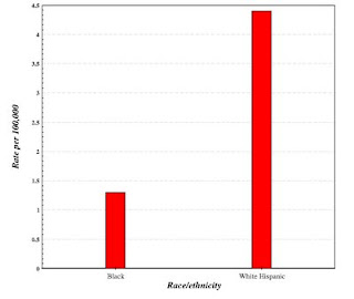
You may be wondering, if I am interested in germ cell tumors, why did I do a query on tumors of the testes. I did this because the SEER interface does not allow me to do a query on specific types of germ cell tumors or of any specific testicular tumor. I know that most testicular tumors are germ cell tumors, so I settled, figuring that if there were a difference in the incidence of germ cell tumors in the Hispanic and the African-American populations, it would show up in the query.
And that's what happened. The SEER output demonstrated that white Hispanics had a much higher incidence of testicular tumors (and, presumably, testicular germ cell tumors) than the African-American population.
If I want to find the ratio for specific tumors, I need to do a little more work. A simple Perl script produced the following list:
The left column is the ratio of cases per total population of white Hispanics divided by the same ratio for African-Americans. The second column is the number of cases, the third column is the average age of cases, and the final column is the ICD-0 term for the neoplasm.
We see (column 1) that white Hispanics have a higher case ratio for every type of germ cell tumor in males (regardless of site).
This tells us a few things. First, that all of the germ cell tumors are related to each other by more than histogenesis (cell of origin). They must have a relationship that extends to causation and development. Second, it tells us that the relatively high level of occurrence of germ cell tumors in white Hispanics is not just a fluke occurring in one cancer of one particular site. It is a consistent phenomenon that extends to several different related tumors and their histologic variants.
I should stress that germ cell tumors are rare, even in the Hispanic population. We are discussing relative rates of uncommon tumors among different ethnicities. An individual's risk of developing a germ cell tumor is low, regardless of ethnicity.
For Perl and Ruby programmers, methods and scripts for using SEER and other publicly available biomedical databases, are described in detail in my prior books:
Perl Programming for Medicine and Biology
Ruby Programming for Medicine and Biology
More information on cancer is available in my recently published book, Neoplasms: Principles of Development and Diversity.
- © 2008 Jules Berman
key words: neoplasms, cancer, neoplasia, precancer, tumor, tumour, tumors, tumours, neoplasm, carcinogenesis, carcinogens, tumor genetics
As requested in the SEER Public-Use Agreement, the SEER citation is included here:
"Surveillance, Epidemiology, and End Results (SEER) Program (www.seer.cancer.gov) Limited-Use Data (1973-2005), National Cancer Institute, DCCPS, Surveillance Research Program, Cancer Statistics Branch, released April 2008, based on the November 2007 submission."
As with all of my scripts, lists, web sites, and blog entries, the following disclaimer applies. This material is provided by its creator, Jules J. Berman, "as is", without warranty of any kind, expressed or implied, including but not limited to the warranties of merchantability, fitness for a particular purpose and noninfringement. in no event shall the author or copyright holder be liable for any claim, damages or other liability, whether in an action of contract, tort or otherwise, arising from, out of or in connection with the material or the use or other dealings in the material.
In a prior blog, I showed how to do a global search through the SEER public use neoplasm incidence files, producing a list of every neoplastic entity captured in the approx 3.5 million SEER cases, and categorizing them by ethnicity, average age of occurrence, number of occurrences, and other data culled from the master data sets.
Reviewing the output, I found that the Hispanic and White populations had a greater number of occurrences of germ cell tumors (an uncommon type of tumor) than the African-American population.
I went to the SEER site and used SEER's public query engine to see if this observation could be verified.
The SEER query site is:
http://seer.cancer.gov/canques/index.html
All queries begin here. I looked for tumor in males, in testes, comparing Hispanic non-whites with African-Americans. A simple interface permits these selections.
The SEER interface produces a list of your input parameters.

The same interface produces a bar chart of your results:

You may be wondering, if I am interested in germ cell tumors, why did I do a query on tumors of the testes. I did this because the SEER interface does not allow me to do a query on specific types of germ cell tumors or of any specific testicular tumor. I know that most testicular tumors are germ cell tumors, so I settled, figuring that if there were a difference in the incidence of germ cell tumors in the Hispanic and the African-American populations, it would show up in the query.
And that's what happened. The SEER output demonstrated that white Hispanics had a much higher incidence of testicular tumors (and, presumably, testicular germ cell tumors) than the African-American population.
If I want to find the ratio for specific tumors, I need to do a little more work. A simple Perl script produced the following list:
2.031 0173 026 germinoma
2.756 0005 038 intratubular malignant germ cells *
2.756 0005 025 malignant teratoma, undifferentiated type *
3.409 0104 019 teratoma, malignant nos
3.478 1125 035 seminoma nos
4.452 0054 036 seminoma, anaplastic type
4.757 0157 028 teratocarcinoma
5.053 0060 027 choriocarcinoma combined with teratoma
5.168 0018 050 spermatocytic seminoma *
5.523 0299 028 embryonal carcinoma nos
5.548 0363 028 mixed germ cell tumor
6.389 0028 027 germ cell tumor, nonseminomatous

* Cases with asterisks have too few cases (second column)
for any significance
The left column is the ratio of cases per total population of white Hispanics divided by the same ratio for African-Americans. The second column is the number of cases, the third column is the average age of cases, and the final column is the ICD-0 term for the neoplasm.
We see (column 1) that white Hispanics have a higher case ratio for every type of germ cell tumor in males (regardless of site).
This tells us a few things. First, that all of the germ cell tumors are related to each other by more than histogenesis (cell of origin). They must have a relationship that extends to causation and development. Second, it tells us that the relatively high level of occurrence of germ cell tumors in white Hispanics is not just a fluke occurring in one cancer of one particular site. It is a consistent phenomenon that extends to several different related tumors and their histologic variants.
I should stress that germ cell tumors are rare, even in the Hispanic population. We are discussing relative rates of uncommon tumors among different ethnicities. An individual's risk of developing a germ cell tumor is low, regardless of ethnicity.
For Perl and Ruby programmers, methods and scripts for using SEER and other publicly available biomedical databases, are described in detail in my prior books:
Perl Programming for Medicine and Biology
Ruby Programming for Medicine and Biology
More information on cancer is available in my recently published book, Neoplasms: Principles of Development and Diversity.
- © 2008 Jules Berman
key words: neoplasms, cancer, neoplasia, precancer, tumor, tumour, tumors, tumours, neoplasm, carcinogenesis, carcinogens, tumor genetics
As requested in the SEER Public-Use Agreement, the SEER citation is included here:
"Surveillance, Epidemiology, and End Results (SEER) Program (www.seer.cancer.gov) Limited-Use Data (1973-2005), National Cancer Institute, DCCPS, Surveillance Research Program, Cancer Statistics Branch, released April 2008, based on the November 2007 submission."
As with all of my scripts, lists, web sites, and blog entries, the following disclaimer applies. This material is provided by its creator, Jules J. Berman, "as is", without warranty of any kind, expressed or implied, including but not limited to the warranties of merchantability, fitness for a particular purpose and noninfringement. in no event shall the author or copyright holder be liable for any claim, damages or other liability, whether in an action of contract, tort or otherwise, arising from, out of or in connection with the material or the use or other dealings in the material.
Sunday, November 16, 2008
Using SEER Public Use Data: 3
Earlier today, I provided a blog with a large list of named neoplasms, listing ratios of occurrences in the African-American and white U.S. populations
http://julesberman.blogspot.com/2008/11/using-seer-public-use-data-2.html
For space limitions, the full name of the neoplasms was truncated.
I've prepared a separate web page, containing the full-length names of neoplasms, along with additional commentary.
http://www.julesberman.info/seerwhbl.htm
In June, 2014, my book, entitled Rare Diseases and Orphan Drugs: Keys to Understanding and Treating the Common Diseases was published by Elsevier. The book builds the argument that our best chance of curing the common diseases will come from studying and curing the rare diseases.
I urge you to read more about my book. There's a generous preview of the book at the Google Books site. If you like the book, please request your librarian to purchase a copy of this book for your library or reading room.
- Jules J. Berman, Ph.D., M.D.
Using SEER Public Use Data: 2
Continuing my previous blog on the SEER Public Use Data files, here is a listing of the ratios of cancer cases, of different types, occurring in the white population and the African-American population in the U.S.
As a rough guide to interpreting the data, neoplasms found in the top third of the list occur disproportionately often in the African-American population. Neoplasms found in the bottom third of the list occur disproportionately often in the white population. Neoplasms in the middle third occur in similar proportions in both sets.
The left hand column is the ratio of occurrence of the tumors in the two populations. Each ratio was calculated as the fraction of cases of the tumor in the white population divided by the fraction of the cases of the tumor in the black population. If the tumor accounted for the same fraction of total cancer cases in the white and black populations, it would have a ratio of 1. It is important to note that we cannot simply find the ratio of the the tumor's occurrence in the white and black populations (because there are many more whites than blacks).
You might be wondering why this question (i.e., tumors in blacks vs tumors in whites) has any clinical importance or biological relevance. It's a good question, and it doesn't have an answer that will satisfy everyone. First, the information helps us avoid diagnostic errors. For example, a pathologist should be wary about making a diagnosis of Ewing's tumor or of superficial spreading melanoma in an African American (in whom these tumors seldom occur). Second, the list generates new hypotheses. We notice that germ cell tumors (including seminomas, teratomas and embryonal carcinomas) occur much for frequently in whites than in the black population. Why is this? Is there some gene that contributes to these tumors, that occurs more frequenly in the white population? We cannot ask such questions if we do not have the kinds of observations included in this list. Third, the list can be generated with the Public Use data sets, which contain race/ethnicity data in case records.
In a later blog, I will show the short Perl script that generated the table from the SEER Public Use Data.
The first column is the case occurrence ratio (white/black). The second column is the number of cases, for each tumor type, in the SEER public use data sets. Tumors occurring with under 40 cases were excluded. The third column is the average age of patients with the neoplasm. The fourth column is the ICD-O(international classification of diseases - oncology) neoplasm term, truncated for space limitation.
In the next few blogs, I'll explain the clinical significance of this and other demo projects, and I'll describe the free, open source, techniques that I used to extract and compile the data. Afterwards, I'll show you how we can drill into the data to refine the questions we can ask, so that we can draw conclusions that can stand up to critical inspection.
For Perl and Ruby programmers, methods and scripts for using SEER and other publicly available biomedical databases, are described in detail in my prior books:
Perl Programming for Medicine and Biology
Ruby Programming for Medicine and Biology
More information on cancer is available in my recently published book, Neoplasms.
- © 2008 Jules Berman
key words: neoplasms, cancer, neoplasia, precancer, tumor, tumour, tumors, tumours, neoplasm, carcinogenesis, carcinogens, tumor genetics
As specified in the SEER Data Agreement, the citation for the SEER data is as follows:
"Surveillance, Epidemiology, and End Results (SEER) Program (www.seer.cancer.gov) Limited-Use Data (1973-2005), National Cancer Institute, DCCPS, Surveillance Research Program, Cancer Statistics Branch, released April 2008, based on the November 2007 submission."
As with all of my scripts, lists, web sites, and blog entries, the following disclaimer applies. This material is provided by its creator, Jules J. Berman, "as is", without warranty of any kind, expressed or implied, including but not limited to the warranties of merchantability, fitness for a particular purpose and noninfringement. in no event shall the author or copyright holder be liable for any claim, damages or other liability, whether in an action of contract, tort or otherwise, arising from, out of or in connection with the material or the use or other dealings in the material.
As a rough guide to interpreting the data, neoplasms found in the top third of the list occur disproportionately often in the African-American population. Neoplasms found in the bottom third of the list occur disproportionately often in the white population. Neoplasms in the middle third occur in similar proportions in both sets.
The left hand column is the ratio of occurrence of the tumors in the two populations. Each ratio was calculated as the fraction of cases of the tumor in the white population divided by the fraction of the cases of the tumor in the black population. If the tumor accounted for the same fraction of total cancer cases in the white and black populations, it would have a ratio of 1. It is important to note that we cannot simply find the ratio of the the tumor's occurrence in the white and black populations (because there are many more whites than blacks).
You might be wondering why this question (i.e., tumors in blacks vs tumors in whites) has any clinical importance or biological relevance. It's a good question, and it doesn't have an answer that will satisfy everyone. First, the information helps us avoid diagnostic errors. For example, a pathologist should be wary about making a diagnosis of Ewing's tumor or of superficial spreading melanoma in an African American (in whom these tumors seldom occur). Second, the list generates new hypotheses. We notice that germ cell tumors (including seminomas, teratomas and embryonal carcinomas) occur much for frequently in whites than in the black population. Why is this? Is there some gene that contributes to these tumors, that occurs more frequenly in the white population? We cannot ask such questions if we do not have the kinds of observations included in this list. Third, the list can be generated with the Public Use data sets, which contain race/ethnicity data in case records.
In a later blog, I will show the short Perl script that generated the table from the SEER Public Use Data.
The first column is the case occurrence ratio (white/black). The second column is the number of cases, for each tumor type, in the SEER public use data sets. Tumors occurring with under 40 cases were excluded. The third column is the average age of patients with the neoplasm. The fourth column is the ICD-O(international classification of diseases - oncology) neoplasm term, truncated for space limitation.
White/Black No. cases Avg age ICD-0 Diagnosis
  00.113    0000040   040     pigmented dermatofibrosarcoma protuberans
  00.190    0000126   060     adult t-cell leukemia/lymphoma (htlv-1 pos.)
  00.225    0000056   068     granular cell tumor, malignant
  00.227    0000057   061     collecting duct carcinoma
  00.238    0000045   056     thymoma, type ab, malignant
  00.263    0000089   050     ameloblastoma, malignant
  00.271    0000053   053     hypereosinophilic syndrome
  00.314    0000116   051     gastrinoma, malignant
  00.314    0000042   053     odontogenic tumor, malignant
  00.372    0000103   026     alveolar soft part sarcoma
  00.394    0000120   055     atypical medullary carcinoma
  00.405    0000335   051     medullary carcinoma with lymphoid stroma
  00.415    0000053   038     craniopharyngioma
  00.416    0000481   039     hodgkin lymph., nodular lymphocyte predom.
  00.419    0000115   030     precursor t-cell lymphoblastic lymphoma
  00.428    0000200   065     plasma cell leukemia
  00.439    0000832   031     choriocarcinoma
  00.441    0000050   002     infantile fibrosarcoma
  00.443    0001263   062     gastrointestinal stromal sarcoma
  00.444    0003634   042     dermatofibrosarcoma nos
  00.447    0000040   040     sertoli-leydig cell tumor, poorly differentiated
  00.448    0000106   038     mesenchymal chondrosarcoma
  00.461    0001225   050     pituitary adenoma, nos
  00.461    0000042   066     prolymphocytic leukemia, t-cell type
  00.464    0000959   055     thymoma, malignant
  00.484    0000091   059     polymorphous low grade adenocarcinoma
  00.485    0019957   064     hepatocellular carcinoma nos
  00.494    0000780   053     granulosa cell tumor, malignant
  00.496    0000227   028     chondroblastic osteosarcoma
  00.501    0000779   067     mesodermal mixed tumor
  00.501    0023109   063     squamous cell carcinoma, keratinizing type nos
  00.502    0002766   072     adenocarcinoma, intestinal type
  00.504    0036429   069     multiple myeloma
  00.507    0000641   001     retinoblastoma nos
  00.510    0000482   045     sq. cell carcinoma, keratinizing, nos, in situ
  00.511    0001466   005     nephroblastoma nos
  00.514    0000140   056     pleomorphic rhabdomyosarcoma
  00.515    0000303   061     metaplastic carcinoma, nos
  00.517    0000117   024     precursor t-cell lymphoblastic leukemia
  00.520    0000337   018     alveolar rhabdomyosarcoma
  00.531    0000105   065     adenocarcinoma with neuroendocrine differen.
  00.533    0000117   051     paraganglioma, malignant
  00.541    0003377   060     mycosis fungoides
  00.544    0003088   068     mullerian mixed tumor
  00.550    0000756   014     embryonal rhabdomyosarcoma
  00.551    0007534   053     medullary carcinoma nos
  00.553    0003575   070     tumor cells, malignant
  00.553    0000617   060     renal cell carcinoma, chromophobe type
  00.553    0000299   060     squamous cell carcinoma, small cell, nonkeratining
  00.554    0000514   067     intracystic carcinoma, nos
  00.558    0000118   028     parosteal osteosarcoma
  00.565    0000070   055     juvenile carcinoma of the breast
  00.570    0057571   038     carcinoma in situ nos
  00.571    0000196   049     pheochromocytoma, malignant
  00.573    0000156   033     synovial sarcoma, biphasic type
  00.577    0000054   058     malignant myoepithelioma
  00.584    0000114   059     atypical meningioma
  00.585    0000041   000     retinoblastoma, differentiated type
  00.586    0000486   060     epithelioid leiomyosarcoma
  00.596    0000183   065     superficial spreading adenocarcinoma
  00.597    0001267   066     carcinoma, diffuse type
  00.598    0000906   061     meningioma, malignant
  00.602    0000191   058     stromal sarcoma, nos
  00.613    0001094   065     intraductal papillary adenocarcinoma with invasion
  00.617    0000135   063     composite carcinoid
  00.626    0000043   002     retinoblastoma, undifferentiated type
  00.627    0000118   037     giant cell tumor of bone, malignant
  00.629    0001848   033     osteosarcoma nos
  00.630    0016028   060     carcinoid tumor, malignant
  00.631    0000090   024     malignant rhabdoid tumor
  00.633    0000202   058     myeloid sarcoma
  00.634    0000403   057     adenosarcoma
  00.634    0000229   058     meningotheliomatous meningioma
  00.637    0000757   064     papillary squamous cell carcinoma
  00.637    0008089   058     squamous cell carcinoma, large cell, nonkeratinizing
  00.638    0003873   044     squamous cell carcinoma, microinvasive
  00.639    0000069   021     ganglioglioma
  00.639    0000118   062     epithelial-myoepithelial carcinoma
  00.641    0000586   069     chronic myeloproliferative disease, nos
  00.643    0001258   052     fibrosarcoma nos
  00.654    0000884   051     lymphoepithelial carcinoma
  00.658    0014107   041     kaposi's sarcoma
  00.661    0000690   047     neurofibrosarcoma
  00.662    0000514   065     pleomorphic carcinoma
  00.667    0247826   064     squamous cell carcinoma nos
  00.673    0000082   059     thymic carcinoma, nos
  00.673    0000047   047     leydig cell tumor, malignant
  00.673    0000050   066     adenocarc. in situ in mult. adenomatous polyps
  00.674    0000179   049     mesenchymoma, malignant
  00.678    0014878   068     non-small cell carcinoma
  00.681    0000890   054     anaplastic large cell lymphoma, t-cell and null cell
  00.682    0002723   065     meningioma nos
  00.682    0000861   042     hodgkin's disease, lymphocytic predominance
  00.683    0000114   033     fibroblastic osteosarcoma
  00.685    0000282   042     epithelioid cell sarcoma
  00.685    0000507   019     primitive neuroectodermal tumor
  00.685    0000123   040     clear cell sarcoma of tendons and aponeuroses
  00.686    0000087   037     small cell sarcoma
  00.686    0000334   063     giant cell sarcoma (except of bone m9250/3)
  00.687    0008070   059     leiomyosarcoma nos
  00.688    0003285   057     inflammatory carcinoma
  00.690    0000555   037     synovial sarcoma nos
  00.692    0000058   052     hodgkin's disease, lymphocytic depletion, diffuse fib
  00.693    0001762   004     neuroblastoma nos
  00.693    0000257   048     megakaryocytic leukemia
  00.697    0000939   063     plasmacytoma, extramedullary
  00.702    0003124   059     sarcoma nos
  00.703    0004558   059     infiltr. duct mixed with other types of carcinoma, in
  00.706    0002264   068     carcinosarcoma nos
  00.707    0001065   064     giant cell carcinoma
  00.707    0000053   026     telangiectatic osteosarcoma
  00.707    0000084   014     langerhans cell histiocytosis, disseminated
  00.712    0002593   062     infiltr. duct mixed with other types of carcinoma
  00.727    0000049   022     precursor b-cell lymphoblastic lymphoma
  00.728    0010967   065     signet ring cell carcinoma
  00.735    0000723   064     apocrine adenocarcinoma
  00.745    0000903   055     endometrial stromal sarcoma
  00.750    0001267   060     mature t-cell lymphoma, nos
  00.751    0000085   050     acute myeloid leukemia, t(8;21)(q22;q22)
  00.755    0000869   063     plasma cell tumor, malignant
  00.755    0000194   048     endometrial stromal sarcoma, low grade
  00.761    0010254   064     carcinoma, undifferentiated type nos
  00.762    0003162   062     noninfiltrating intraductal papillary adenocarcinoma
  00.763    0000934   052     cystosarcoma phyllodes, malignant
  00.768    0000295   068     pseudosarcomatous carcinoma
  00.773    0011520   064     acinar cell carcinoma
  00.774    0000584   067     squamous cell carcinoma, spindle cell type
  00.782    0000493   062     basaloid squamous cell carcinoma
  00.785    0029592   065     large cell carcinoma nos
  00.786    0000787   066     spindle cell carcinoma
  00.786    0000277   063     combined hepatocellular carcinoma and cholangiocarcino
  00.791    0025451   067     mucin-producing adenocarcinoma
  00.794    0001025   060     spindle cell sarcoma
  00.796    0006891   056     comedocarcinoma nos
  00.796    0000258   062     renal cell carcinoma, sarcomatoid
  00.800    0000123   065     brenner tumor, malignant
  00.805    0016555   068     adenocarcinoma in tubulovillous adenoma
  00.808    0000051   035     prolactinoma
  00.809    0005831   067     adenocarcinoma in situ in tubulovillous adenoma
  00.811    0024527   041     squamous intraepithelial neoplasia, grade iii
  00.812    0000234   058     malignant tumor, small cell type
  00.812    0000248   052     chronic myelogenous leukemia, bcr/abl positive
  00.814    0004050   050     follicular adenocarcinoma nos
  00.824    0000856   065     adenocarcinoma with mixed subtypes
  00.824    0000060   016     choroid plexus papilloma, malignant
  00.828    0000046   053     fascial fibrosarcoma
  00.829    0000843   058     intraductal micropapillary carcinoma
  00.831    0000947   064     granular cell carcinoma
  00.833    0000079   044     papillary cystadenoma, borderline malignancy (c56.9)
  00.839    0000324   061     acute myeloid leukemia, minimal differentiation
  00.841    0000584   020     endodermal sinus tumor
  00.845    0000722   050     neurilemmoma, malignant
  00.845    0000557   067     combined small cell carcinoma
  00.846    0002217   061     paget's disease and infiltrating duct carcinoma of brea
  00.851    0000505   043     rhabdomyosarcoma nos
  00.851    0011193   063     adenosquamous carcinoma
  00.852    0000265   053     fibromyxosarcoma
  00.854    0000139   022     ependymoma, anaplastic type
  00.858    0000066   057     thymoma, type b1, malignant
  00.858    0000045   037     mediastinal large b-cell lymphoma
  00.858    0000388   037     sq. cell carcinoma, lg. cell, non-ker., in situ
  00.859    0000416   038     hodgkin lymphoma, nod. scler., grade 1
  00.861    0000517   047     follicular adenocarcinoma, well differentiated type
  00.868    0000052   065     eccrine adenocarcinoma
  00.868    0063370   038     squamous cell carcinoma in situ nos
  00.870    0037543   064     renal cell carcinoma
  00.871    0001654   066     linitis plastica
  00.873    0001021   060     liposarcoma, well differentiated type
  00.874    0000328   055     adenocarcinoid tumor
  00.874    0000382   061     mixed tumor, malignant nos
  00.877    0002754   056     intraductal and lobular in situ carcinoma
  00.877    0000141   050     follicular adenocarcinoma, trabecular type
  00.878    0000100   061     papillary squamous cell carcinoma, non-invasive
  00.880    0001242   023     teratoma, malignant nos
  00.880    0000080   071     refract. anemia with excess blasts in transformation
  00.883    0005106   064     neuroendocrine carcinoma
  00.884    0182616   072     carcinoma nos
  00.884    0003757   054     mucoepidermoid carcinoma
  00.885    0000179   051     adenocarcinoma in adenomatous polyposis coli
  00.897    0000100   019     desmoplastic medulloblastoma
  00.900    0000690   028     germinoma
  00.904    1021940   068     adenocarcinoma nos
  00.907    0056558   076     neoplasm, malignant
  00.909    0000055   020     clear cell sarcoma of kidney
  00.920    0003319   058     adenoid cystic carcinoma
  00.921    0000082   032     neuroepithelioma nos
  00.923    0010280   061     chronic myeloid leukemia
  00.927    0003078   048     hodgkin's disease nos
  00.927    0000963   035     precursor cell lymphoblastic lymphoma, nos
  00.930    0000165   067     bronchiolo-alveolar carcinoma, non-mucinous
  00.933    0000884   062     acral lentiginous melanoma, malig.
  00.934    0000308   007     ganglioneuroblastoma
  00.937    0000265   055     mucocarcinoid tumor, malignant
  00.937    0000403   066     large cell neuroendocrine carcinoma
  00.946    0000089   057     myxosarcoma
  00.958    0000820   073     refractory anemia
  00.959    0013199   064     malignant lymphoma nos
  00.963    0000315   066     malignant tumor, fusiform cell type
  00.966    0000086   045     hemangioblastoma
  00.967    0000855   057     islet cell carcinoma
  00.968    0001163   013     medulloblastoma nos
  00.968    0000191   052     myxoid chondrosarcoma
  00.970    0001140   047     acute promyelocytic leukemia
  00.972    0000096   056     myxoid leiomyosarcoma
  00.972    0000093   025     pleomorphic xanthoastrocytoma
  00.972    0000087   064     acute panmyelosis with myelofibrosis
  00.974    0002700   050     bowen's disease
  00.981    0000080   063     basal cell adenocarcinoma
  00.982    0000125   065     nodular hidradenoma, malignant
  00.989    0000114   067     sezary's disease
  00.989    0000115   051     round cell liposarcoma
  00.992    0035630   059     intraductal carcinoma, noninfiltrating nos
  00.993    0000358   051     hemangiopericytoma, malignant
  00.996    0003164   064     scirrhous adenocarcinoma
  00.997    0003337   046     glioma, malignant
  01.003    0001045   060     cutaneous t-cell lymphoma, nos
  01.027    0001609   037     burkitt lymphoma, nos
  01.028    0001015   067     lymphoid leukemia nos
  01.032    0000104   048     epithelioid hemangioendothelioma, malignant
  01.036    0000303   004     hepatoblastoma
  01.036    0000998   065     essential thrombocythemia
  01.040    0000390   066     alveolar adenocarcinoma
  01.047    0000094   035     protoplasmic astrocytoma
  01.050    0016754   069     adenocarcinoma in villous adenoma
  01.050    0001041   049     serous cystadenoma, borderline malignancy (c56.9)
  01.058    0000548   038     hodgkin lymphoma, nod. scler., cellular phase
  01.059    0001545   017     pilocytic astrocytoma (c71._) 9421/1
  01.061    0008163   067     adenocarcinoma in situ in adenomatous polyp
  01.065    0000815   066     erythroleukemia
  01.066    0001669   066     small cell carcinoma, intermediate cell
  01.067    0000248   064     giant cell and spindle cell carcinoma
  01.071    0001388   034     ependymoma nos
  01.072    0000624   066     papillary carcinoma in situ
  01.075    0004414   046     hodgkin's disease, mixed cellularity
  01.089    0001323   062     liposarcoma nos
  01.092    0000142   054     mesonephroma, malignant
  01.092    0000212   067     bronchiolo-alveolar carcinoma, mucinous
  01.093    0001167   053     myxoid liposarcoma
  01.100    0050173   066     small cell carcinoma nos
  01.103    0004446   065     carcinoma, anaplastic type nos
  01.111    0000159   052     hemangioendothelioma, malignant
  01.116    0002422   074     myelodysplastic syndrome, nos
  01.117    0333623   061     infiltrating duct carcinoma
  01.120    0000405   064     pleomorphic liposarcoma
  01.132    0005812   062     fibrous histiocytoma, malignant
  01.132    0003638   065     marginal zone b-cell lymphoma, nos
  01.134    0004672   069     adenocarcinoma in situ in villous adenoma
  01.138    0000145   057     mixed type liposarcoma
  01.144    0000084   063     psammomatous meningioma
  01.149    0000166   060     carcinoid tumor, argentaffin, malignant
  01.154    0000096   039     hepatocellular carcinoma, fibrolamellar
  01.162    0000655   074     refractory anemia with sideroblasts
  01.166    0000151   064     adenocarcinoma in mult. adenomatous polyps
  01.166    0001603   048     serous papillary cystic tumor of borderline malignancy
  01.169    0011933   033     hodgkin lymphoma, nodular sclerosis, nos
  01.171    0018131   061     acute myeloid leukemia
  01.173    0000287   063     dedifferentiated liposarcoma
  01.173    0009679   058     comedocarcinoma, noninfiltrating
  01.175    0027965   059     papillary adenocarcinoma nos
  01.178    0000091   045     papillary carcinoma, encapsulated
  01.184    0001275   064     polycythemia vera
  01.189    0020880   068     adenocarcinoma in adenomatous polyp
  01.189    0000827   021     precursor b-cell lymphoblastic leukemia
  01.190    0000215   046     follicular carcinoma, minimally invasive
  01.191    0000461   066     basaloid carcinoma
  01.196    0000178   037     synovial sarcoma, spindle cell type
  01.196    0000362   069     myelosclerosis with myeloid metaplasia
  01.197    0001872   069     myeloid leukemia nos
  01.198    0045778   068     mucinous adenocarcinoma
  01.200    0002839   059     cribriform carcinoma in situ
  01.206    0000516   050     papillary microcarcinoma
  01.208    0000435   036     hodgkin lymphoma, nod. scler., grade 2
  01.220    0000165   061     carcinoma in pleomorphic adenoma
  01.223    0000614   060     acute myeloid leukemia with maturation
  01.234    0001092   062     mixed cell adenocarcinoma
  01.243    0009222   023     precursor cell lymphoblastic leukemia, nos
  01.249    0020557   061     clear cell adenocarcinoma nos
  01.254    0011881   066     bronchiolo-alveolar adenocarcinoma
  01.255    0000115   044     adenocarcinoma, endocervical type
  01.257    0001270   066     ml, lymphoplasmacytic
  01.262    0000144   056     transitional meningioma
  01.263    0004480   058     ml, large b-cell, diffuse, immunoblastic, nos
  01.265    0010260   046     papillary and follicular adenocarcinoma
  01.279    0000172   050     giant cell glioblastoma
  01.287    0000057   055     chromophobe carcinoma
  01.287    0000127   066     neoplasm, uncertain whether benign or malignant
  01.288    0001435   058     oxyphilic adenocarcinoma
  01.295    0000880   065     cribriform carcinoma
  01.300    0000485   056     papillary mucinous cystadenocarcinoma
  01.302    0000149   034     peripheral neuroectodermal tumor
  01.312    0004610   069     cholangiocarcinoma
  01.312    0002006   061     acute myelomonocytic leukemia
  01.313    0000626   056     hodgkin's disease, lymphocytic depletion nos
  01.320    0015063   062     papillary serous cystadenocarcinoma
  01.326    0000241   066     angioimmunoblastic t-cell lymphoma
  01.329    0000117   054     nk/t-cell lymphoma, nasal and nasal-type
  01.336    0000758   068     villous adenocarcinoma
  01.341    0000761   051     adrenal cortical carcinoma
  01.348    0001142   063     cystadenocarcinoma nos
  01.356    0000618   066     paget's disease, mammary
  01.362    0009635   067     ml, small b lymphocytic, nos
  01.363    0003731   065     acute leukemia nos
  01.364    0001353   063     hemangiosarcoma
  01.371    0008335   047     astrocytoma nos
  01.372    0001358   064     cloacogenic carcinoma
  01.376    0010414   054     lobular carcinoma in situ
  01.391    0021695   061     infiltrating duct and lobular carcinoma
  01.397    0017866   064     oat cell carcinoma
  01.407    0002708   063     serous surface papillary carcinoma
  01.414    0000054   065     hepatocellular carcinoma, clear cell type
  01.425    0000285   046     burkitt's tumor
  01.425    0003428   056     mucinous cystadenocarcinoma nos
  01.437    0004682   062     serous cystadenocarcinoma nos
  01.445    0000301   070     prolymphocytic leukemia, nos
  01.451    0000327   067     acute myeloid leuk. with multilineage dysplasia
  01.454    0000156   069     basosquamous carcinoma
  01.455    0037088   063     ml, large b-cell, diffuse
  01.481    0000050   059     solitary fibrous tumor, malignant
  01.487    0001358   063     paget disease and intraductal ca.
  01.487    0001004   050     medullary carcinoma with amyloid stroma
  01.488    0012432   062     malignant lymphoma, non hodgkin's type
  01.491    0004319   065     ml, mixed sm. and lg. cell, diffuse
  01.497    0001673   068     verrucous carcinoma nos
  01.512    0002581   072     leukemia nos
  01.522    0001770   058     acute monocytic leukemia
  01.530    0001669   060     duct carcinoma in situ, solid type
  01.535    0000086   065     adenoid squamous cell carcinoma
  01.535    0000086   062     therapy-related myelodysplastic syndrome, nos
  01.540    0000079   058     thymoma, type b3, malignant
  01.559    0030328   070     chronic lymphoid leukemia
  01.599    0000111   047     papillary mucinous cystadenoma, borderline malignancy (
  01.605    0000179   067     splenic marginal zone b-cell lymphoma
  01.617    0001798   074     chronic myelomonocytic leukemia, nos
  01.627    0006684   058     adenocarcinoma in situ
  01.632    0000111   055     fibrous meningioma
  01.641    0000074   061     hodgkin's disease, lymphocytic depletion, reticular
  01.650    0000763   072     refractory anemia with excess blasts
  01.651    0022382   050     papillary carcinoma nos
  01.658    0000885   061     infiltrating ductular carcinoma
  01.666    0000118   042     burkitt cell leukemia
  01.692    0001301   059     papillary cystadenocarcinoma nos
  01.721    0050243   070     transitional cell carcinoma nos
  01.733    0002320   048     astrocytoma, anaplastic type
  01.738    0000363   064     sweat gland adenocarcinoma
  01.742    0026705   061     endometrioid carcinoma
  01.777    0000500   048     oligodendroglioma, anaplastic type
  01.796    0034336   064     lobular carcinoma nos
  01.818    0000295   061     glioblastoma with sarcomatous component
  01.829    0004897   068     mesothelioma, malignant
  01.846    0000302   052     esthesioneuroblastoma
  01.863    0001932   051     chondrosarcoma nos
  01.868    0000042   061     adenocarcinoma with apocrine metaplasia
  01.905    0000472   063     infiltrating lobular mixed with other types of carc.
  01.924    0000448   059     acute myeloid leukemia without maturation
  01.931    0001012   040     mixed glioma
  01.935    0000159   049     nonencapsulated sclerosing carcinoma
  01.986    0000128   053     spermatocytic seminoma
  01.987    0001486   049     mucinous cystic tumor of borderline malignancy (c56.9)
  01.994    0002890   066     mantle cell lymphoma
  02.002    0000764   042     fibrillary astrocytoma
  02.020    0000184   067     trabecular adenocarcinoma
  02.020    0000046   069     eccrine poroma, malignant
  02.034    0003345   063     malignant lymphoma, nodular nos
  02.046    0000966   031     dysgerminoma
  02.053    0000070   040     myxopapillary ependymoma
  02.057    0006337   062     tubular adenocarcinoma
  02.088    0000978   052     neurilemmoma nos
  02.090    0000219   038     astroblastoma
  02.119    0003414   062     malignant lymphoma, follicular center cell, noncleaved,
  02.171    0019907   061     glioblastoma nos
  02.222    0000239   063     solid carcinoma nos
  02.229    0001585   071     waldenstrom macroglobulinemia
  02.235    0000940   069     transitional cell carcinoma in situ
  02.242    0000122   071     transitional cell carcinoma, spindle cell type
  02.289    0000077   062     atypical carcinoid tumor
  02.333    0002297   041     oligodendroglioma nos
  02.363    0000263   071     refractory cytopenia with multilineage dysplasia
  02.491    0000089   074     myelodysplastic syndr. with 5q deletion syndrome
  02.525    0000054   068     carcinoma simplex
  02.554    0000461   049     gemistocytic astrocytoma
  02.558    0000083   064     small cell carcinoma, fusiform cell type
  02.592    0000086   056     papillary carcinoma, columnar cell
  02.657    0000408   072     klatskin tumor
  02.666    0000147   056     primary cutan. cd30+ t-cell lymphoprolif. disorder
  02.726    0096537   068     papillary transitional cell carcinoma
  02.790    0005812   062     malignant lymphoma, mixed lymphocytic-histiocytic, nod
  02.891    0000614   055     chordoma
  02.932    0008938   060     malignant lymphoma, follicular center cell, cleaved,
  02.962    0000476   069     fibrous mesothelioma, malignant
  03.094    0002388   030     mixed germ cell tumor
  03.131    0000786   072     basal cell carcinoma nos
  03.245    0002438   069     papillary trans. cell carcinoma, non-invasive
  03.257    0000135   054     malignant mastocytosis
  03.282    0000494   062     skin appendage carcinoma
  03.491    0000261   072     mesothelioma, biphasic type, malignant
  03.570    0000796   072     sebaceous adenocarcinoma
  03.587    0001263   067     epithelioid mesothelioma, malignant
  03.602    0000113   060     queyrat's erythroplasia
  03.602    0000116   065     sclerosing sweat duct carcinoma
  03.694    0002147   058     hairy cell leukemia
  03.695    0004086   059     adenocarcinoma with squamous metaplasia
  03.737    0000042   056     gliomatosis cerebri
  04.040    0000041   064     lymphangiosarcoma
  04.040    0000095   028     germ cell tumor, nonseminomatous
  04.337    0009849   037     seminoma nos
  04.747    0000049   070     osteosarcoma in paget's disease of bone
  04.955    0001717   028     teratocarcinoma
  05.050    0000057   048     ac. myelomonocytic leuk. w abn. mar. eosinophils
  05.529    0003184   030     embryonal carcinoma nos
  05.611    0001088   019     ewing's sarcoma
  05.824    0000556   028     choriocarcinoma combined with teratoma
  06.019    0000627   038     seminoma, anaplastic type
  06.565    0000401   060     epithelioid cell melanoma
  08.055    0000811   071     paget's disease, extramammary (except paget's of bone)
  08.206    0001716   074     merkel cell carcinoma
  10.024    0000412   056     malignant melanoma, regressing
  10.706    0000110   052     precancerous melanosis nos
  10.891    0000661   061     mixed epithel. & spindle cell melanoma
  12.623    0048315   056     malignant melanoma nos
  18.888    0000764   060     amelanotic melanoma
  19.367    0000783   066     desmoplastic melanoma, malignant
  21.009    0001281   062     spindle cell melanoma nos
  24.492    0008688   060     nodular melanoma
  29.788    0007272   069     malignant melanoma in hutchinson's melanotic freckle
  31.608    0021999   057     melanoma in situ
  38.463    0040929   052     superficial spreading melanoma
  38.730    0018652   068     hutchinson's melanotic freckle
  47.472    0000477   058     spindle cell melanoma, type b
  62.046    0004547   051     superficial spreading melanoma, in situ
In the next few blogs, I'll explain the clinical significance of this and other demo projects, and I'll describe the free, open source, techniques that I used to extract and compile the data. Afterwards, I'll show you how we can drill into the data to refine the questions we can ask, so that we can draw conclusions that can stand up to critical inspection.
For Perl and Ruby programmers, methods and scripts for using SEER and other publicly available biomedical databases, are described in detail in my prior books:
Perl Programming for Medicine and Biology
Ruby Programming for Medicine and Biology
More information on cancer is available in my recently published book, Neoplasms.
- © 2008 Jules Berman
key words: neoplasms, cancer, neoplasia, precancer, tumor, tumour, tumors, tumours, neoplasm, carcinogenesis, carcinogens, tumor genetics
As specified in the SEER Data Agreement, the citation for the SEER data is as follows:
"Surveillance, Epidemiology, and End Results (SEER) Program (www.seer.cancer.gov) Limited-Use Data (1973-2005), National Cancer Institute, DCCPS, Surveillance Research Program, Cancer Statistics Branch, released April 2008, based on the November 2007 submission."
As with all of my scripts, lists, web sites, and blog entries, the following disclaimer applies. This material is provided by its creator, Jules J. Berman, "as is", without warranty of any kind, expressed or implied, including but not limited to the warranties of merchantability, fitness for a particular purpose and noninfringement. in no event shall the author or copyright holder be liable for any claim, damages or other liability, whether in an action of contract, tort or otherwise, arising from, out of or in connection with the material or the use or other dealings in the material.
Friday, November 14, 2008
Using SEER Public Use Data: 1
SEER is the U.S. National Cancer Institute's Surveillance, Epidemiology and End Results program. It is an amazing resource for information about the cancers that occur in the U.S. One of the products of SEER is the Public Use dataset, which contains de-identified records on over 3.5 million cancers that have occurred between 1973 and 2005.
When you have 3.5 million cancer cases to study, you can draw certain types of inferences that could not possibly be made with the data accumulated at a single medical institution.
I thought I would do a series of blogs, extended over the next few weeks or months, showing how the SEER dataset can be analyzed, the kinds of hypotheses and discoveries that can be made by studying the database, and the kinds of things that you can do when you combine SEER data with data from other publicly available resources.
Each SEER record is a cancer case, described by a series of 258 (mostly) numbers, in byte-assigned positions, described by a data dictionary document. When you have the byte locations for the data dictionary entries, you can easily write a short script (I like to use Perl, Ruby, or Python) that can extract and compile data any way you wish.
For example, the following list is a compilation of all of the diagnoses (that occur at least 10 times in the data set), sorted by the age of the person at the time of diagnosis, accompanied by the number of cases and the cumulative fraction of cases accounted for, and by the diagnosis (truncated for reasons of space).
As specified in the Limited-Use Data Agreement, the citation for the SEER data
is as follows:
Once we have the columned data, we can easily produce a graphic that represents the salient features we would like to emphasize.

In the next few blogs, I'll explain the clinical significance of this little demo project, and I'll describe the free, open source, techniques that I used to extract and compile the data. Afterwards, I'll show you how we can drill into the data to refine the questions we can ask, so that we can draw conclusions that can stand up to critical inspection.
For Perl and Ruby programmers, methods and scripts for using SEER and other publicly available biomedical databases, are described in detail in my prior books:
Perl Programming for Medicine and Biology
Ruby Programming for Medicine and Biology
- © 2008 Jules Berman
As specified in the SEER Data Agreement, the citation for the SEER data is as follows:
"Surveillance, Epidemiology, and End Results (SEER) Program (www.seer.cancer.gov) Limited-Use Data (1973-2005), National Cancer Institute, DCCPS, Surveillance Research Program, Cancer Statistics Branch, released April 2008, based on the November 2007 submission."
As with all of my scripts, lists, web sites, and blog entries, the following disclaimer applies. This material is provided by its creator, Jules J. Berman, "as is", without warranty of any kind, expressed or implied, including but not limited to the warranties of merchantability, fitness for a particular purpose and noninfringement. in no event shall the author or copyright holder be liable for any claim, damages or other liability, whether in an action of contract, tort or otherwise, arising from, out of or in connection with the material or the use or other dealings in the material.
In June, 2014, my book, entitled Rare Diseases and Orphan Drugs: Keys to Understanding and Treating the Common Diseases was published by Elsevier. The book builds the argument that our best chance of curing the common diseases will come from studying and curing the rare diseases.
I urge you to read more about my book. There's a generous preview of the book at the Google Books site. If you like the book, please request your librarian to purchase a copy of this book for your library or reading room.
When you have 3.5 million cancer cases to study, you can draw certain types of inferences that could not possibly be made with the data accumulated at a single medical institution.
I thought I would do a series of blogs, extended over the next few weeks or months, showing how the SEER dataset can be analyzed, the kinds of hypotheses and discoveries that can be made by studying the database, and the kinds of things that you can do when you combine SEER data with data from other publicly available resources.
Each SEER record is a cancer case, described by a series of 258 (mostly) numbers, in byte-assigned positions, described by a data dictionary document. When you have the byte locations for the data dictionary entries, you can easily write a short script (I like to use Perl, Ruby, or Python) that can extract and compile data any way you wish.
For example, the following list is a compilation of all of the diagnoses (that occur at least 10 times in the data set), sorted by the age of the person at the time of diagnosis, accompanied by the number of cases and the cumulative fraction of cases accounted for, and by the diagnosis (truncated for reasons of space).
Age Number  Cumu-         Name
yrs  of     lat-          of
    cases   ive           Neoplasm
-------------------------------------------------
000 0000041 0.000 retinoblastoma, differentiated
001 0000641 0.000 retinoblastoma nos
002 0000050 0.000 infantile fibrosarcoma
002 0000016 0.000 juvenile myelomonocytic leukemi
002 0000021 0.000 atypical teratoid/rhabdoid tumo
002 0000043 0.000 retinoblastoma, undifferentiate
004 0000303 0.000 hepatoblastoma
004 0001762 0.000 neuroblastoma nos
004 0000012 0.000 medulloepithelioma nos
005 0001466 0.001 nephroblastoma nos
007 0000308 0.001 ganglioneuroblastoma
011 0000013 0.001 subependymal giant cell astrocy
012 0000010 0.001 pancreatoblastoma
013 0001163 0.001 medulloblastoma nos
013 0000018 0.001 neurofibromatosis nos
014 0000756 0.001 embryonal rhabdomyosarcoma
014 0000084 0.001 langerhans cell histiocytosis,
016 0000060 0.001 choroid plexus papilloma, malig
017 0001545 0.002 pilocytic astrocytoma (c71._) 9
018 0000031 0.002 embryonal sarcoma
018 0000337 0.002 alveolar rhabdomyosarcoma
019 0001088 0.002 ewing's sarcoma
019 0000100 0.002 desmoplastic medulloblastoma
019 0000507 0.002 primitive neuroectodermal tumor
020 0000584 0.003 endodermal sinus tumor
020 0000055 0.003 clear cell sarcoma of kidney
021 0000069 0.003 ganglioglioma
021 0000827 0.003 precursor b-cell lymphoblastic
022 0000139 0.003 ependymoma, anaplastic type
022 0000025 0.003 dysembryoplastic neuroepithelia
022 0000049 0.003 precursor b-cell lymphoblastic
023 0001242 0.003 teratoma, malignant nos
023 0000021 0.003 choroid plexus papilloma nos
023 0009222 0.006 precursor cell lymphoblastic le
024 0000098 0.006 pineoblastoma
024 0000090 0.006 malignant rhabdoid tumor
024 0000117 0.006 precursor t-cell lymphoblastic
025 0000011 0.006 periosteal osteosarcoma
025 0000023 0.006 mixed type rhabdomyosarcoma
025 0000093 0.006 pleomorphic xanthoastrocytoma
026 0000103 0.006 alveolar soft part sarcoma
026 0000019 0.006 chondroblastoma, malignant
026 0000053 0.006 telangiectatic osteosarcoma
027 0000011 0.006 spindle cell rhabdomyosarcoma
027 0000039 0.006 desmoplastic small round cell t
028 0000690 0.006 germinoma
028 0001717 0.007 teratocarcinoma
028 0000118 0.007 parosteal osteosarcoma
028 0000227 0.007 chondroblastic osteosarcoma
028 0000095 0.007 germ cell tumor, nonseminomatou
028 0000556 0.007 choriocarcinoma combined with t
029 0000023 0.007 ganglioglioma, anaplastic
029 0000019 0.007 intratubular malignant germ cel
029 0000021 0.007 malignant placental site tropho
030 0002388 0.008 mixed germ cell tumor
030 0003184 0.009 embryonal carcinoma nos
030 0000115 0.009 precursor t-cell lymphoblastic
031 0000966 0.009 dysgerminoma
031 0000832 0.009 choriocarcinoma
031 0000021 0.009 centrol neurocytoma
032 0000016 0.009 lipoma nos
032 0000082 0.009 neuroepithelioma nos
032 0000027 0.009 adamantinomatous craniopharyngi
032 0000019 0.009 malignant teratoma, undifferent
033 0001848 0.010 osteosarcoma nos
033 0000114 0.010 fibroblastic osteosarcoma
033 0000030 0.010 adamantinoma of long bones
033 0000156 0.010 synovial sarcoma, biphasic type
033 0011933 0.013 hodgkin lymphoma, nodular scler
034 0001388 0.014 ependymoma nos
034 0000149 0.014 peripheral neuroectodermal tumo
035 0000051 0.014 prolactinoma
035 0000094 0.014 protoplasmic astrocytoma
035 0000963 0.014 precursor cell lymphoblastic ly
036 0000435 0.014 hodgkin lymphoma, nod. scler.,
037 0009849 0.017 seminoma nos
037 0000087 0.017 small cell sarcoma
037 0000010 0.017 acidophil carcinoma
037 0000555 0.017 synovial sarcoma nos
037 0001609 0.017 burkitt lymphoma, nos
037 0000045 0.017 mediastinal large b-cell lympho
037 0000178 0.017 synovial sarcoma, spindle cell
037 0000118 0.018 giant cell tumor of bone, malig
037 0000388 0.018 sq. cell carcinoma, lg. cell, n
038 0000219 0.018 astroblastoma
038 0000053 0.018 craniopharyngioma
038 0057571 0.034 carcinoma in situ nos
038 0000016 0.034 androblastoma, malignant
038 0000627 0.034 seminoma, anaplastic type
038 0000106 0.034 mesenchymal chondrosarcoma
038 0063370 0.052 squamous cell carcinoma in situ
038 0000416 0.052 hodgkin lymphoma, nod. scler.,
038 0000548 0.052 hodgkin lymphoma, nod. scler.,
039 0000019 0.052 neurofibroma nos
039 0000026 0.052 sertoli cell carcinoma
039 0000096 0.052 hepatocellular carcinoma, fibro
039 0000021 0.052 hepatosplenic gamma-delta cell
039 0000481 0.052 hodgkin lymph., nodular lymphoc
039 0000024 0.052 mpnst with rhabdomyoblastic dif
040 0001012 0.053 mixed glioma
040 0000038 0.053 cavernous hemangioma
040 0000070 0.053 myxopapillary ependymoma
040 0000040 0.053 pigmented dermatofibrosarcoma p
040 0000123 0.053 clear cell sarcoma of tendons a
040 0000040 0.053 sertoli-leydig cell tumor, poor
041 0014107 0.057 kaposi's sarcoma
041 0002297 0.057 oligodendroglioma nos
041 0000014 0.057 spongioblastoma polare
041 0000010 0.057 papillary carcinoma, oxyphilic
041 0024527 0.064 squamous intraepithelial neopla
042 0000024 0.064 pulmonary blastoma
042 0000118 0.064 burkitt cell leukemia
042 0000764 0.065 fibrillary astrocytoma
042 0003634 0.066 dermatofibrosarcoma nos
042 0000282 0.066 epithelioid cell sarcoma
042 0000861 0.066 hodgkin's disease, lymphocytic
043 0000036 0.066 hemangioma nos
043 0000505 0.066 rhabdomyosarcoma nos
043 0000019 0.066 solid pseudopapillary carcinoma
044 0000015 0.066 papillary meningioma
044 0000013 0.066 papillary meningioma 9538/3
044 0000017 0.066 juxtacortical chondrosarcoma
044 0000115 0.066 adenocarcinoma, endocervical ty
044 0003873 0.067 squamous cell carcinoma, microi
044 0000079 0.067 papillary cystadenoma, borderli
045 0000086 0.067 hemangioblastoma
045 0000037 0.067 oligodendroblastoma
045 0000016 0.067 struma ovarii, malignant
045 0000029 0.067 undifferentiated sarcoma
045 0000010 0.067 clear cell chondrosarcoma
045 0000011 0.067 carotid body tumor, malignant
045 0000091 0.067 papillary carcinoma, encapsulat
045 0000035 0.067 extra-adrenal paraganglioma, ma
045 0000482 0.067 sq. cell carcinoma, keratinizin
046 0000285 0.068 burkitt's tumor
046 0003337 0.069 glioma, malignant
046 0000024 0.069 periosteal fibrosarcoma
046 0004414 0.070 hodgkin's disease, mixed cellul
046 0010260 0.073 papillary and follicular adenoc
046 0000215 0.073 follicular carcinoma, minimally
047 0008335 0.075 astrocytoma nos
047 0000690 0.075 neurofibrosarcoma
047 0000047 0.075 leydig cell tumor, malignant
047 0001140 0.076 acute promyelocytic leukemia
047 0000163 0.076 malignant melanoma in giant pig
047 0000517 0.076 follicular adenocarcinoma, well
047 0000111 0.076 papillary mucinous cystadenoma,
047 0000084 0.076 squamous cell carcinoma in situ
048 0003078 0.077 hodgkin's disease nos
048 0000257 0.077 megakaryocytic leukemia
048 0000039 0.077 ovarian stromal tumor, mal.
048 0002320 0.077 astrocytoma, anaplastic type
048 0000500 0.078 oligodendroglioma, anaplastic t
048 0000194 0.078 endometrial stromal sarcoma, lo
048 0000104 0.078 epithelioid hemangioendotheliom
048 0000057 0.078 ac. myelomonocytic leuk. w abn.
048 0001603 0.078 serous papillary cystic tumor o
049 0000033 0.078 subependymal glioma
049 0000179 0.078 mesenchymoma, malignant
049 0000461 0.078 gemistocytic astrocytoma
049 0000012 0.078 primary effusion lymphoma
049 0000196 0.078 pheochromocytoma, malignant
049 0000159 0.078 nonencapsulated sclerosing carc
049 0000034 0.078 dermoid cyst with malignant tra
049 0001041 0.079 serous cystadenoma, borderline
049 0001486 0.079 mucinous cystic tumor of border
050 0002700 0.080 bowen's disease
050 0000036 0.080 hodgkin's granuloma
050 0000012 0.080 chromophobe adenoma
050 0001225 0.080 pituitary adenoma, nos
050 0000722 0.080 neurilemmoma, malignant
050 0000172 0.081 giant cell glioblastoma
050 0022382 0.087 papillary carcinoma nos
050 0000089 0.087 ameloblastoma, malignant
050 0000516 0.087 papillary microcarcinoma
050 0000034 0.087 acute biphenotypic leukemia
050 0004050 0.088 follicular adenocarcinoma nos
050 0000017 0.088 mixed medullary-papillary carci
050 0001004 0.088 medullary carcinoma with amyloi
050 0000085 0.088 acute myeloid leukemia, t(8;21)
050 0000030 0.088 mucinous cystadenocarcinoma, no
050 0000029 0.088 mucinous adenocarcinoma, endoce
050 0000141 0.089 follicular adenocarcinoma, trab
051 0001932 0.089 chondrosarcoma nos
051 0000116 0.089 gastrinoma, malignant
051 0000115 0.089 round cell liposarcoma
051 0000117 0.089 paraganglioma, malignant
051 0000761 0.089 adrenal cortical carcinoma
051 0000884 0.090 lymphoepithelial carcinoma
051 0000358 0.090 hemangiopericytoma, malignant
051 0004547 0.091 superficial spreading melanoma,
051 0000335 0.091 medullary carcinoma with lympho
051 0000026 0.091 malignant giant cell tumor of s
051 0000179 0.091 adenocarcinoma in adenomatous p
052 0000043 0.091 myosarcoma
052 0000153 0.091 adenoma nos
052 0000978 0.091 neurilemmoma nos
052 0001258 0.092 fibrosarcoma nos
052 0000032 0.092 histiocytic sarcoma
052 0000191 0.092 myxoid chondrosarcoma
052 0000302 0.092 esthesioneuroblastoma
052 0000110 0.092 precancerous melanosis nos
052 0040929 0.104 superficial spreading melanoma
052 0000159 0.104 hemangioendothelioma, malignant
052 0000934 0.104 cystosarcoma phyllodes, maligna
052 0000031 0.104 malignant melanoma in precancer
052 0000248 0.104 chronic myelogenous leukemia, b
052 0000058 0.104 hodgkin's disease, lymphocytic
053 0000097 0.104 neoplasm, benign
053 0000265 0.104 fibromyxosarcoma
053 0001167 0.104 myxoid liposarcoma
053 0000046 0.104 fascial fibrosarcoma
053 0000027 0.104 blue nevus, malignant
053 0000128 0.104 spermatocytic seminoma
053 0007534 0.107 medullary carcinoma nos
053 0000053 0.107 hypereosinophilic syndrome
053 0000042 0.107 odontogenic tumor, malignant
053 0000780 0.107 granulosa cell tumor, malignant
053 0000350 0.107 malignant melanoma in junctiona
054 0000032 0.107 neuroma nos
054 0000025 0.107 balloon cell melanoma
054 0000135 0.107 malignant mastocytosis
054 0000142 0.107 mesonephroma, malignant
054 0003757 0.108 mucoepidermoid carcinoma
054 0010414 0.111 lobular carcinoma in situ
054 0000029 0.111 thymoma, type b2, malignant
054 0000117 0.111 nk/t-cell lymphoma, nasal and n
054 0000890 0.111 anaplastic large cell lymphoma,
055 0000614 0.111 chordoma
055 0000023 0.111 angiomyosarcoma
055 0000111 0.111 fibrous meningioma
055 0000959 0.112 thymoma, malignant
055 0000328 0.112 adenocarcinoid tumor
055 0000057 0.112 chromophobe carcinoma
055 0000032 0.112 megakaryocytic myelosis
055 0000903 0.112 endometrial stromal sarcoma
055 0000120 0.112 atypical medullary carcinoma
055 0000265 0.112 mucocarcinoid tumor, malignant
055 0000070 0.112 juvenile carcinoma of the breas
055 0000029 0.112 adenocarcinoma in situ in famil
056 0000042 0.112 gliomatosis cerebri
056 0006891 0.114 comedocarcinoma nos
056 0000028 0.114 theca cell carcinoma
056 0000096 0.114 myxoid leiomyosarcoma
056 0048315 0.128 malignant melanoma nos
056 0000030 0.128 angiomatous meningioma
056 0000144 0.128 transitional meningioma
056 0000045 0.128 thymoma, type ab, malignant
056 0000140 0.128 pleomorphic rhabdomyosarcoma
056 0000412 0.128 malignant melanoma, regressing
056 0003428 0.129 mucinous cystadenocarcinoma nos
056 0000086 0.129 papillary carcinoma, columnar c
056 0000485 0.129 papillary mucinous cystadenocar
056 0002754 0.130 intraductal and lobular in situ
056 0000626 0.130 hodgkin's disease, lymphocytic
056 0000039 0.130 endometrioid adenocarcinoma, se
056 0000147 0.130 primary cutan. cd30+ t-cell lym
057 0000089 0.130 myxosarcoma
057 0000403 0.130 adenosarcoma
057 0021999 0.137 melanoma in situ
057 0000855 0.137 islet cell carcinoma
057 0000145 0.137 mixed type liposarcoma
057 0003285 0.138 inflammatory carcinoma
057 0000066 0.138 thymoma, type b1, malignant
057 0000047 0.138 spindle cell melanoma, type a
057 0000026 0.138 subcutaneous panniculitis-like
058 0000011 0.138 vipoma
058 0000202 0.138 myeloid sarcoma
058 0002147 0.138 hairy cell leukemia
058 0000191 0.139 stromal sarcoma, nos
058 0000038 0.139 insulinoma, malignant
058 0000032 0.139 glucagonoma, malignant
058 0006684 0.140 adenocarcinoma in situ
058 0001435 0.141 oxyphilic adenocarcinoma
058 0001770 0.141 acute monocytic leukemia
058 0000054 0.141 malignant myoepithelioma
058 0003319 0.142 adenoid cystic carcinoma
058 0000079 0.142 thymoma, type b3, malignant
058 0000477 0.142 spindle cell melanoma, type b
058 0000229 0.142 meningotheliomatous meningioma
058 0000234 0.143 malignant tumor, small cell typ
058 0009679 0.145 comedocarcinoma, noninfiltratin
058 0000207 0.145 cyst-associated renal cell carc
058 0000843 0.146 intraductal micropapillary carc
058 0004480 0.147 ml, large b-cell, diffuse, immu
058 0008089 0.149 squamous cell carcinoma, large
059 0003124 0.150 sarcoma nos
059 0008070 0.152 leiomyosarcoma nos
059 0000114 0.152 atypical meningioma
059 0000082 0.152 thymic carcinoma, nos
059 0000016 0.152 aggressive nk-cell leukemia
059 0002839 0.153 cribriform carcinoma in situ
059 0027965 0.161 papillary adenocarcinoma nos
059 0000018 0.161 malignant eccrine spiradenoma
059 0001301 0.161 papillary cystadenocarcinoma no
059 0000050 0.161 solitary fibrous tumor, maligna
059 0000091 0.161 polymorphous low grade adenocar
059 0004086 0.163 adenocarcinoma with squamous me
059 0000448 0.163 acute myeloid leukemia without
059 0035630 0.173 intraductal carcinoma, noninfil
059 0000022 0.173 acute myeloid leukemia, 11q23 a
059 0000020 0.173 mixed islet cell and exocrine a
059 0004558 0.174 infiltr. duct mixed with other
060 0008688 0.176 nodular melanoma
060 0000030 0.176 insular carcinoma
060 0003377 0.177 mycosis fungoides
060 0000764 0.178 amelanotic melanoma
060 0001025 0.178 spindle cell sarcoma
060 0000113 0.178 queyrat's erythroplasia
060 0000401 0.178 epithelioid cell melanoma
060 0016028 0.183 carcinoid tumor, malignant
060 0000486 0.183 epithelioid leiomyosarcoma
060 0001267 0.183 mature t-cell lymphoma, nos
060 0001045 0.183 cutaneous t-cell lymphoma, nos
060 0000020 0.183 carcinosarcoma, embryonal type
060 0001669 0.184 duct carcinoma in situ, solid t
060 0001021 0.184 liposarcoma, well differentiate
060 0000014 0.184 granulosa cell-theca cell tumor
060 0000614 0.184 acute myeloid leukemia with mat
060 0000617 0.184 renal cell carcinoma, chromopho
060 0000166 0.185 carcinoid tumor, argentaffin, m
060 0000126 0.185 adult t-cell leukemia/lymphoma
060 0000299 0.185 squamous cell carcinoma, small
060 0008938 0.187 malignant lymphoma, follicular
061 0019907 0.193 glioblastoma nos
061 0000906 0.193 meningioma, malignant
061 0000015 0.193 epithelioma, malignant
061 0026705 0.201 endometrioid carcinoma
061 0018131 0.206 acute myeloid leukemia
061 0010280 0.209 chronic myeloid leukemia
061 0000026 0.209 polygonal cell carcinoma
061 0000057 0.209 collecting duct carcinoma
061 0000303 0.209 metaplastic carcinoma, nos
061 0000382 0.209 mixed tumor, malignant nos
061 0333623 0.303 infiltrating duct carcinoma
061 0002006 0.303 acute myelomonocytic leukemia
061 0020557 0.309 clear cell adenocarcinoma nos
061 0000885 0.309 infiltrating ductular carcinoma
061 0000165 0.309 carcinoma in pleomorphic adenom
061 0000661 0.310 mixed epithel. & spindle cell m
061 0000295 0.310 glioblastoma with sarcomatous c
061 0000042 0.310 adenocarcinoma with apocrine me
061 0021695 0.316 infiltrating duct and lobular c
061 0000324 0.316 acute myeloid leukemia, minimal
061 0000100 0.316 papillary squamous cell carcino
061 0000012 0.316 endometrioid adenocarcinoma, ci
061 0000074 0.316 hodgkin's disease, lymphocytic
061 0002217 0.317 paget's disease and infiltratin
062 0001323 0.317 liposarcoma nos
062 0006337 0.319 tubular adenocarcinoma
062 0000014 0.319 heavy chain disease, nos
062 0000077 0.319 atypical carcinoid tumor
062 0000494 0.319 skin appendage carcinoma
062 0001092 0.319 mixed cell adenocarcinoma
062 0001281 0.320 spindle cell melanoma nos
062 0004682 0.321 serous cystadenocarcinoma nos
062 0005812 0.323 fibrous histiocytoma, malignant
062 0001263 0.323 gastrointestinal stromal sarcom
062 0000106 0.323 malignant tumor, giant cell typ
062 0000493 0.323 basaloid squamous cell carcinom
062 0000258 0.323 renal cell carcinoma, sarcomato
062 0000118 0.323 epithelial-myoepithelial carcin
062 0000884 0.323 acral lentiginous melanoma, mal
062 0015063 0.328 papillary serous cystadenocarci
062 0000039 0.328 endometrioid adenofibroma, mali
062 0012432 0.331 malignant lymphoma, non hodgkin
062 0000037 0.331 adenocarcinoma with spindle cel
062 0000086 0.331 therapy-related myelodysplastic
062 0002593 0.332 infiltr. duct mixed with other
062 0003162 0.333 noninfiltrating intraductal pap
062 0005812 0.334 malignant lymphoma, mixed lymph
062 0003414 0.335 malignant lymphoma, follicular
063 0001353 0.336 hemangiosarcoma
063 0000023 0.336 hodgkin's sarcoma
063 0000024 0.336 meningiomatosis nos
063 0000239 0.336 solid carcinoma nos
063 0000135 0.336 composite carcinoid
063 0001142 0.336 cystadenocarcinoma nos
063 0000023 0.336 schneiderian carcinoma
063 0011193 0.339 adenosquamous carcinoma
063 0000084 0.339 psammomatous meningioma
063 0037088 0.350 ml, large b-cell, diffuse
063 0000080 0.350 basal cell adenocarcinoma
063 0000016 0.350 intestinal t-cell lymphoma
063 0000287 0.350 dedifferentiated liposarcoma
063 0000939 0.350 plasmacytoma, extramedullary
063 0000869 0.350 plasma cell tumor, malignant
063 0003345 0.351 malignant lymphoma, nodular nos
063 0001358 0.352 paget disease and intraductal c
063 0002708 0.353 serous surface papillary carcin
063 0000020 0.353 squamous cell carcinoma, clear
063 0000012 0.353 basal cell carcinoma, fibroepit
063 0000334 0.353 giant cell sarcoma (except of b
063 0023109 0.359 squamous cell carcinoma, kerati
063 0000472 0.359 infiltrating lobular mixed with
063 0000277 0.359 combined hepatocellular carcino
064 0001275 0.360 polycythemia vera
064 0000041 0.360 lymphangiosarcoma
064 0017866 0.365 oat cell carcinoma
064 0037543 0.375 renal cell carcinoma
064 0001065 0.376 giant cell carcinoma
064 0011520 0.379 acinar cell carcinoma
064 0001358 0.379 cloacogenic carcinoma
064 0034336 0.389 lobular carcinoma nos
064 0013199 0.393 malignant lymphoma nos
064 0000947 0.393 granular cell carcinoma
064 0000405 0.393 pleomorphic liposarcoma
064 0000723 0.393 apocrine adenocarcinoma
064 0003164 0.394 scirrhous adenocarcinoma
064 0005106 0.396 neuroendocrine carcinoma
064 0000363 0.396 sweat gland adenocarcinoma
064 0247826 0.465 squamous cell carcinoma nos
064 0019957 0.471 hepatocellular carcinoma nos
064 0000757 0.471 papillary squamous cell carcino
064 0010254 0.474 carcinoma, undifferentiated typ
064 0000087 0.474 acute panmyelosis with myelofib
064 0000248 0.474 giant cell and spindle cell car
064 0000083 0.474 small cell carcinoma, fusiform
064 0000151 0.474 adenocarcinoma in mult. adenoma
064 0000018 0.474 atypical chronic myeloid leuk.,
064 0000026 0.474 adenocarcinoma with cartilagino
065 0002723 0.475 meningioma nos
065 0003731 0.476 acute leukemia nos
065 0000880 0.476 cribriform carcinoma
065 0000200 0.477 plasma cell leukemia
065 0000514 0.477 pleomorphic carcinoma
065 0000052 0.477 eccrine adenocarcinoma
065 0029592 0.485 large cell carcinoma nos
065 0000123 0.485 brenner tumor, malignant
065 0000998 0.485 essential thrombocythemia
065 0000016 0.485 ceruminous adenocarcinoma
065 0010967 0.488 signet ring cell carcinoma
065 0000125 0.488 nodular hidradenoma, malignant
065 0004446 0.490 carcinoma, anaplastic type nos
065 0000086 0.490 adenoid squamous cell carcinoma
065 0000116 0.490 sclerosing sweat duct carcinoma
065 0000856 0.490 adenocarcinoma with mixed subty
065 0003638 0.491 marginal zone b-cell lymphoma,
065 0004319 0.492 ml, mixed sm. and lg. cell, dif
065 0000183 0.492 superficial spreading adenocarc
065 0000054 0.492 hepatocellular carcinoma, clear
065 0000020 0.492 composite hodgkin and non-hodgk
065 0000105 0.492 adenocarcinoma with neuroendocr
065 0001094 0.493 intraductal papillary adenocarc
066 0000815 0.493 erythroleukemia
066 0001654 0.493 linitis plastica
066 0000461 0.493 basaloid carcinoma
066 0002890 0.494 mantle cell lymphoma
066 0001270 0.495 ml, lymphoplasmacytic
066 0000787 0.495 spindle cell carcinoma
066 0001267 0.495 carcinoma, diffuse type
066 0000390 0.495 alveolar adenocarcinoma
066 0000618 0.496 paget's disease, mammary
066 0050173 0.510 small cell carcinoma nos
066 0000624 0.510 papillary carcinoma in situ
066 0000783 0.510 desmoplastic melanoma, malignan
066 0011881 0.513 bronchiolo-alveolar adenocarcin
066 0000241 0.513 angioimmunoblastic t-cell lymph
066 0000403 0.514 large cell neuroendocrine carci
066 0000315 0.514 malignant tumor, fusiform cell
066 0000042 0.514 prolymphocytic leukemia, t-cell
066 0001669 0.514 small cell carcinoma, intermedi
066 0000050 0.514 adenocarc. in situ in mult. ade
066 0000127 0.514 neoplasm, uncertain whether ben
066 0000032 0.514 intraductal papillary-mucinous
067 0000114 0.514 sezary's disease
067 0001015 0.515 lymphoid leukemia nos
067 0000779 0.515 mesodermal mixed tumor
067 0000184 0.515 trabecular adenocarcinoma
067 0000514 0.515 intracystic carcinoma, nos
067 0009635 0.518 ml, small b lymphocytic, nos
067 0000557 0.518 combined small cell carcinoma
067 0025451 0.525 mucin-producing adenocarcinoma
067 0000032 0.525 dedifferentiated chondrosarcoma
067 0000016 0.525 immunoproliferative disease, no
067 0001263 0.525 epithelioid mesothelioma, malig
067 0000179 0.525 splenic marginal zone b-cell ly
067 0000212 0.525 bronchiolo-alveolar carcinoma,
067 0000584 0.526 squamous cell carcinoma, spindl
067 0008163 0.528 adenocarcinoma in situ in adeno
067 0000165 0.528 bronchiolo-alveolar carcinoma,
067 0005831 0.530 adenocarcinoma in situ in tubul
067 0000327 0.530 acute myeloid leuk. with multil
067 0000026 0.530 bronch.-alv. carc., mixed mucin
067 0000038 0.530 intraductal papillary-mucinous
068 0000054 0.530 carcinoma simplex
068 0002264 0.530 carcinosarcoma nos
068 1021940 0.818 adenocarcinoma nos
068 0003088 0.819 mullerian mixed tumor
068 0000758 0.819 villous adenocarcinoma
068 0045778 0.832 mucinous adenocarcinoma
068 0004897 0.834 mesothelioma, malignant
068 0001673 0.834 verrucous carcinoma nos
068 0014878 0.838 non-small cell carcinoma
068 0000032 0.838 thymoma, type a, malignant
068 0000295 0.838 pseudosarcomatous carcinoma
068 0000056 0.838 granular cell tumor, malignant
068 0018652 0.844 hutchinson's melanotic freckle
068 0020880 0.849 adenocarcinoma in adenomatous p
068 0000032 0.849 prolymphocytic leukemia, b-cell
068 0096537 0.877 papillary transitional cell car
068 0016555 0.881 adenocarcinoma in tubulovillous
069 0036429 0.892 multiple myeloma
069 0004610 0.893 cholangiocarcinoma
069 0001872 0.893 myeloid leukemia nos
069 0000156 0.893 basosquamous carcinoma
069 0000046 0.893 eccrine poroma, malignant
069 0000010 0.893 clear cell adenocarcinofibroma
069 0000476 0.894 fibrous mesothelioma, malignant
069 0016754 0.898 adenocarcinoma in villous adeno
069 0000940 0.899 transitional cell carcinoma in
069 0000419 0.899 noninfiltrating intracystic car
069 0000362 0.899 myelosclerosis with myeloid met
069 0000586 0.899 chronic myeloproliferative dise
069 0004672 0.900 adenocarcinoma in situ in villo
069 0002438 0.901 papillary trans. cell carcinoma
069 0007272 0.903 malignant melanoma in hutchinso
070 0003575 0.904 tumor cells, malignant
070 0030328 0.913 chronic lymphoid leukemia
070 0000301 0.913 prolymphocytic leukemia, nos
070 0050243 0.927 transitional cell carcinoma nos
070 0000049 0.927 osteosarcoma in paget's disease
071 0001585 0.927 waldenstrom macroglobulinemia
071 0000122 0.927 transitional cell carcinoma, sp
071 0000263 0.927 refractory cytopenia with multi
071 0000080 0.927 refract. anemia with excess bla
071 0000811 0.928 paget's disease, extramammary (
072 0002581 0.928 leukemia nos
072 0182616 0.980 carcinoma nos
072 0000408 0.980 klatskin tumor
072 0000012 0.980 hepatoid adenocarcinoma
072 0000796 0.980 sebaceous adenocarcinoma
072 0000786 0.980 basal cell carcinoma nos
072 0000012 0.980 adenoid basal cell carcinoma
072 0002766 0.981 adenocarcinoma, intestinal type
072 0000015 0.981 multicentric basal cell carcino
072 0000763 0.981 refractory anemia with excess b
072 0000261 0.981 mesothelioma, biphasic type, ma
072 0000028 0.981 transitional cell carcinoma, mi
073 0000820 0.982 refractory anemia
073 0000036 0.982 basal cell carcinoma, nodular
074 0001716 0.982 merkel cell carcinoma
074 0002422 0.983 myelodysplastic syndrome, nos
074 0000655 0.983 refractory anemia with siderobl
074 0001798 0.984 chronic myelomonocytic leukemia
074 0000089 0.984 myelodysplastic syndr. with 5q
076 0056558 1.000 neoplasm, malignant
As specified in the Limited-Use Data Agreement, the citation for the SEER data
is as follows:
   Surveillance, Epidemiology, and End Results (SEER) Program
   (www.seer.cancer.gov) Limited-Use Data (1973-2005), National Cancer
   Institute, DCCPS, Surveillance Research Program, Cancer Statistics
   Branch, released April 2008, based on the November 2007 submission.
Once we have the columned data, we can easily produce a graphic that represents the salient features we would like to emphasize.

In the next few blogs, I'll explain the clinical significance of this little demo project, and I'll describe the free, open source, techniques that I used to extract and compile the data. Afterwards, I'll show you how we can drill into the data to refine the questions we can ask, so that we can draw conclusions that can stand up to critical inspection.
For Perl and Ruby programmers, methods and scripts for using SEER and other publicly available biomedical databases, are described in detail in my prior books:
Perl Programming for Medicine and Biology
Ruby Programming for Medicine and Biology
- © 2008 Jules Berman
As specified in the SEER Data Agreement, the citation for the SEER data is as follows:
"Surveillance, Epidemiology, and End Results (SEER) Program (www.seer.cancer.gov) Limited-Use Data (1973-2005), National Cancer Institute, DCCPS, Surveillance Research Program, Cancer Statistics Branch, released April 2008, based on the November 2007 submission."
As with all of my scripts, lists, web sites, and blog entries, the following disclaimer applies. This material is provided by its creator, Jules J. Berman, "as is", without warranty of any kind, expressed or implied, including but not limited to the warranties of merchantability, fitness for a particular purpose and noninfringement. in no event shall the author or copyright holder be liable for any claim, damages or other liability, whether in an action of contract, tort or otherwise, arising from, out of or in connection with the material or the use or other dealings in the material.
In June, 2014, my book, entitled Rare Diseases and Orphan Drugs: Keys to Understanding and Treating the Common Diseases was published by Elsevier. The book builds the argument that our best chance of curing the common diseases will come from studying and curing the rare diseases.
I urge you to read more about my book. There's a generous preview of the book at the Google Books site. If you like the book, please request your librarian to purchase a copy of this book for your library or reading room.
Subscribe to:
Posts (Atom)