I'm beginning, for this blog, a series of short utility scripts and essays that relate, in one way or another, to the general subject of indexing and data retrieval.
The first entry is a short Perl script (just 18 command lines) that extracts the names (of people) wherever the names may occur within a provided text file. The output consists of an alphabetized list of non-repeating names. The script is so simple that it can be easily be translated into any language that supports regular expressions (regex).
The script is available at:
http://www.julesberman.info/factoids/namesget.htm
Blog readers who are uninterested in indexing and data retrieval may want to visit my two other blogs,
Machiavelli's Laboratory (scientific ethics taught from the perspective on an unethical scientist)
and
Neoplasms (essays on tumor biology)
- © 2010 Jules Berman
key words: indices, indexing, indexes, index, data retrieval, information retrieval, informatics
Wednesday, October 27, 2010
Thursday, October 14, 2010
Germ cell tumor web page available
The recent blog series on germ cell tumors has been packaged into a single web page available at:
http://www.julesberman.info/factoids/germcell.htm
- © 2010 Jules Berman
key words: carcinogenesis, neoplasia, neoplasms, tumor development, tumour development, tumor biology, tumour biology, carcinogenesis
http://www.julesberman.info/factoids/germcell.htm
- © 2010 Jules Berman
key words: carcinogenesis, neoplasia, neoplasms, tumor development, tumour development, tumor biology, tumour biology, carcinogenesis
Monday, October 11, 2010
New blog site on Neoplasms
Readers who are primarily interested in medical informatics may have found the past week's blogs on germ cell tumors to be a bit off-track. I agree. I've created a new blog site, Neoplasms, to cover my interests in tumor biology.
Henceforth, for the Specified Life blog, I'll stick to issues of data retrieval, organization, indexing, annotation, and analysis.
For readers who have been following the germ cell series, you might be interested in a new blog series I'll be starting, on "epigenomic tumors", in the Neoplasms blog site. I'll be starting with a discussion of malignant rhabdoid tumors.
- © 2010 Jules Berman
key words: carcinogenesis, neoplasia, neoplasms, tumor development, tumour development, tumor biology, tumour biology, carcinogenesis
Henceforth, for the Specified Life blog, I'll stick to issues of data retrieval, organization, indexing, annotation, and analysis.
For readers who have been following the germ cell series, you might be interested in a new blog series I'll be starting, on "epigenomic tumors", in the Neoplasms blog site. I'll be starting with a discussion of malignant rhabdoid tumors.
- © 2010 Jules Berman
key words: carcinogenesis, neoplasia, neoplasms, tumor development, tumour development, tumor biology, tumour biology, carcinogenesis
Thursday, October 7, 2010
Germ cell cancers of testes: conclusion
This is the last entry on a series of blogs on germ cell cancers of the testes.
I've added forward and backward links for each of the blogs in the series, so you can visit the first blog in the series and click forward or backward through the sequential entries.
Basically, in this series, we showed, using the SEER public use data files, that there has been a large increase in the incidence of germ cell cancers of the testis in white non-Hispanic males since the first SEER observation year (1973) up to the most recent data year (2007).
Along with the increase in seminomatous germ cell cancers was a lesser but parallel increase in the non-seminomatous germ cell cancers of the testis, when compared in birth cohort populations.
The seminomatous and non-seminomatous germ cell cancers, though derived from very different cell types (germ cells versus embryonic/extra-embryonic primitive cells) develop from the same precanceous lesion (usually intratubular germ cell neoplasia and sometimes gonadoblastoma). Precancerous germ cells are characterized by epigenomic erasure, and this "erased" state seems to allow precancerous germ cells to develop into seminomas or into tumors derived from totipotent stem cells.
Testicular precancers develop from disorders of sex development. The incidence of disorders of sex development, like the incidence of testicular germ cell cancers, has been rising. The cause for the rise of disorders of sex development (and the concomitant rise in testicular germ cell cancers) is unknown. However, the ubiquitous appearance of the platicizer and endocrine disruptor, Bisphenol A, has captured the interest of toxicologists and cancer researchers.
All of these issues were discussed in this completed series of blogs on testicular germ cell cancers.
- © 2010 Jules Berman
key words: carcinogenesis, neoplasia, neoplasms, tumor development, tumour development, germ cell tumor, germ cell tumour, tumor epidemiology, increasing germ cell cancer rates, germ cell cancer, seminomas, seminomatous, non-seminomatous, non-germinomatous, embryonal carcinoma, choriocarcinoma, testis, testes, itgcn, intratubular germ cell neoplasm, plasticizers, endocrine disruptors
I've added forward and backward links for each of the blogs in the series, so you can visit the first blog in the series and click forward or backward through the sequential entries.
Basically, in this series, we showed, using the SEER public use data files, that there has been a large increase in the incidence of germ cell cancers of the testis in white non-Hispanic males since the first SEER observation year (1973) up to the most recent data year (2007).
Along with the increase in seminomatous germ cell cancers was a lesser but parallel increase in the non-seminomatous germ cell cancers of the testis, when compared in birth cohort populations.
The seminomatous and non-seminomatous germ cell cancers, though derived from very different cell types (germ cells versus embryonic/extra-embryonic primitive cells) develop from the same precanceous lesion (usually intratubular germ cell neoplasia and sometimes gonadoblastoma). Precancerous germ cells are characterized by epigenomic erasure, and this "erased" state seems to allow precancerous germ cells to develop into seminomas or into tumors derived from totipotent stem cells.
Testicular precancers develop from disorders of sex development. The incidence of disorders of sex development, like the incidence of testicular germ cell cancers, has been rising. The cause for the rise of disorders of sex development (and the concomitant rise in testicular germ cell cancers) is unknown. However, the ubiquitous appearance of the platicizer and endocrine disruptor, Bisphenol A, has captured the interest of toxicologists and cancer researchers.
All of these issues were discussed in this completed series of blogs on testicular germ cell cancers.
- © 2010 Jules Berman
key words: carcinogenesis, neoplasia, neoplasms, tumor development, tumour development, germ cell tumor, germ cell tumour, tumor epidemiology, increasing germ cell cancer rates, germ cell cancer, seminomas, seminomatous, non-seminomatous, non-germinomatous, embryonal carcinoma, choriocarcinoma, testis, testes, itgcn, intratubular germ cell neoplasm, plasticizers, endocrine disruptors
Wednesday, October 6, 2010
Explaining the rise in testicular germ cell tumors
In yesterday's blog we saw that the rise in testicular cancer rates in white males showed a parallel increase in seminomatous and non-seminomatous germ cell cancers among birth cohorts.
What can explain this increase?
There is one class of conditions that is overwhelmingly associated with the development of germ cell tumors of the testis: disorders of sex development of the testes.[1] Among the conditions within this general group are testicular dysgenesis, testicular feminization (insensitivity to androgens), and cryptorchidism. Disorders of sex development of the testis raise the incidence of intratubular germ cell neoplasia or of gonadoblastoma, both of which are testicular precancers.
As you might expect, along with the observed increase in testicular germ cell cancers in white males, there has been an observed increase in the incidence of disorders of sex development in the same population. [1,2] These disorders are characterized by a retardation in the maturation of primordial germ cells, along with an apparent mitotic over-stimulation of these same cells: leading to a proliferative, precancerous condition.
Though there is no proof at the moment, we might expect that males who develop testicular germ cell cancer who have clinically normal testes, may harbor small foci of [clinically unobserved] germ cell proliferative lesions.
What has caused the increased incidence of disorders of sex development in the testes? We don't know, but we have a candidate: the ubiquitous plasticizer and endocrine disruptor, Bisphenol A.
Bisphenol A is a synthetic estrogen used in the process of manufacturing plastics, and has been detected in the serum, milk, saliva, urine, and amniotic fluid of humans.[3] Because we get our daily dose of Bisphenol A from plastic bottles, one would expect that the levels of Bisphenol A in our blood would have increased steadily over the past several decades [coinciding with our increased dependence of plastic food and drink containers]. You might also expect that if Bisphenol A produced testicular cancers, you would see the largest increases in incidence among the wealthiest populations in the most industrialized nations [as we do].
Can we assume that Bisphenol A is causing the rise of incidence of testicular germ cell cancers? Absolutely not. All of the evidence, so far, is very weak (if it can be called evidence at all!). Still, nobody would suggest that Bisphenol A has much to recommend itself as a healthy addition to our diets. It seems prudent to try to limit our exposure to this compound when feasible, particularly among infants and pregnant women.
1. Pleskacova J, Hersmus R, Oosterhuis JW, Setyawati BA, Faradz SM, Cools M, Wolffenbuttel KP, Lebl J, Drop SL, Looijenga LH. Tumor Risk in Disorders of Sex Development. Sex Dev 4:259-269, 2010.
2. Skakkebaek NE, Rajpert-De Meyts E, Jorgensen N, Main KM, Leffers H, Andersson AM, Juul A, Jensen TK, Toppari J. Testicular cancer trends as 'whistle blowers' of testicular developmental problems in populations. Int J Androl 30:198-204, 2007.
3. Bouskine A, Nebout M, Brucker-Davis F, Benahmed M, Fenichel P. Low Doses of Bisphenol A Promote Human Seminoma Cell Proliferation by Activating PKA and PKG via a Membrane G-Protein-Coupled Estrogen Receptor. Environ Health Perspect 117:1053-1058, 2009.
This series of blogs has drawn heavily from the public use SEER data sets produced by the U.S. National Cancer Institute's Surveillance, Epidemiology and End Results project. In the next blog, I'll discuss how this data can be obtained and used by the science-minded public.
Jump to Tomorrow's blog
- © 2010 Jules Berman
key words: carcinogenesis, neoplasia, neoplasms, tumor development, tumour development, germ cell tumor, germ cell tumour, tumor epidemiology, increasing germ cell cancer rates, germ cell cancer, seminomas, seminomatous, non-seminomatous, non-germinomatous, embryonal carcinoma, choriocarcinoma, testis, testes, itgcn, intratubular germ cell neoplasm, plasticizers, endocrine disruptors
About my book, Precancer: The Beginning and the End of Cancer. Nearly every type of cancer passes through a precancer phase, during which it cannot metastasize or invade other tissues. While medicine is not always successful in treating or curing advanced stages of cancers, recent advances in our understanding of carcinogenesis have helped us to develop strategies to prevent, diagnose, and treat many cancers at the precancer stage. Research in this field is escalating rapidly as the evidence increasingly shows that the number of annual cancer deaths could be drastically reduced through the effective treatment and cure of precancer lesions. This book begins by explaining why it has been so difficult to cure cancers, followed by a review of precancer biology, with descriptions of the most common precancer lesions. The final chapters provide practical socio-political and medical goals for precancer treatment, including discussions of the economics and politics of treating precancers.

I urge you to read more about this important topic. Google Books has provided a generous preview of this book.
What can explain this increase?
There is one class of conditions that is overwhelmingly associated with the development of germ cell tumors of the testis: disorders of sex development of the testes.[1] Among the conditions within this general group are testicular dysgenesis, testicular feminization (insensitivity to androgens), and cryptorchidism. Disorders of sex development of the testis raise the incidence of intratubular germ cell neoplasia or of gonadoblastoma, both of which are testicular precancers.
As you might expect, along with the observed increase in testicular germ cell cancers in white males, there has been an observed increase in the incidence of disorders of sex development in the same population. [1,2] These disorders are characterized by a retardation in the maturation of primordial germ cells, along with an apparent mitotic over-stimulation of these same cells: leading to a proliferative, precancerous condition.
Though there is no proof at the moment, we might expect that males who develop testicular germ cell cancer who have clinically normal testes, may harbor small foci of [clinically unobserved] germ cell proliferative lesions.
What has caused the increased incidence of disorders of sex development in the testes? We don't know, but we have a candidate: the ubiquitous plasticizer and endocrine disruptor, Bisphenol A.
Bisphenol A is a synthetic estrogen used in the process of manufacturing plastics, and has been detected in the serum, milk, saliva, urine, and amniotic fluid of humans.[3] Because we get our daily dose of Bisphenol A from plastic bottles, one would expect that the levels of Bisphenol A in our blood would have increased steadily over the past several decades [coinciding with our increased dependence of plastic food and drink containers]. You might also expect that if Bisphenol A produced testicular cancers, you would see the largest increases in incidence among the wealthiest populations in the most industrialized nations [as we do].
Can we assume that Bisphenol A is causing the rise of incidence of testicular germ cell cancers? Absolutely not. All of the evidence, so far, is very weak (if it can be called evidence at all!). Still, nobody would suggest that Bisphenol A has much to recommend itself as a healthy addition to our diets. It seems prudent to try to limit our exposure to this compound when feasible, particularly among infants and pregnant women.
1. Pleskacova J, Hersmus R, Oosterhuis JW, Setyawati BA, Faradz SM, Cools M, Wolffenbuttel KP, Lebl J, Drop SL, Looijenga LH. Tumor Risk in Disorders of Sex Development. Sex Dev 4:259-269, 2010.
2. Skakkebaek NE, Rajpert-De Meyts E, Jorgensen N, Main KM, Leffers H, Andersson AM, Juul A, Jensen TK, Toppari J. Testicular cancer trends as 'whistle blowers' of testicular developmental problems in populations. Int J Androl 30:198-204, 2007.
3. Bouskine A, Nebout M, Brucker-Davis F, Benahmed M, Fenichel P. Low Doses of Bisphenol A Promote Human Seminoma Cell Proliferation by Activating PKA and PKG via a Membrane G-Protein-Coupled Estrogen Receptor. Environ Health Perspect 117:1053-1058, 2009.
This series of blogs has drawn heavily from the public use SEER data sets produced by the U.S. National Cancer Institute's Surveillance, Epidemiology and End Results project. In the next blog, I'll discuss how this data can be obtained and used by the science-minded public.
Jump to Tomorrow's blog
- © 2010 Jules Berman
key words: carcinogenesis, neoplasia, neoplasms, tumor development, tumour development, germ cell tumor, germ cell tumour, tumor epidemiology, increasing germ cell cancer rates, germ cell cancer, seminomas, seminomatous, non-seminomatous, non-germinomatous, embryonal carcinoma, choriocarcinoma, testis, testes, itgcn, intratubular germ cell neoplasm, plasticizers, endocrine disruptors
About my book, Precancer: The Beginning and the End of Cancer. Nearly every type of cancer passes through a precancer phase, during which it cannot metastasize or invade other tissues. While medicine is not always successful in treating or curing advanced stages of cancers, recent advances in our understanding of carcinogenesis have helped us to develop strategies to prevent, diagnose, and treat many cancers at the precancer stage. Research in this field is escalating rapidly as the evidence increasingly shows that the number of annual cancer deaths could be drastically reduced through the effective treatment and cure of precancer lesions. This book begins by explaining why it has been so difficult to cure cancers, followed by a review of precancer biology, with descriptions of the most common precancer lesions. The final chapters provide practical socio-political and medical goals for precancer treatment, including discussions of the economics and politics of treating precancers.
I urge you to read more about this important topic. Google Books has provided a generous preview of this book.


Tuesday, October 5, 2010
Explaining the germ cell cancer rates
In yesterday's blog we explained how the precancer of testicular germ cell tumors, intratubular germ cell neoplasia, gives rise to seminomas (differentiated germinomatous lineage) and non-seminomas (tumors of pluripotent progenitor cells that are not of germ cell lineage).
In the first blog of this series on germ cell tumors , we noted that the increase in occurrences of seminomas has outpaced the occurrences of the nonseminomatous germ cell tumors.
Here is a graph, produced from the SEER public use data sets, of the crude occurrences of seminoma and non-seminoma testicular germ cell tumors, in white males, since 1973.

The light blue bars are the seminomas, and the maroon bars are the non-seminomatous germ cell tumors of the testes. Since 1973, the seminomas increased from a number much lower than the occurrences of the non-seminomatous germ cell tumors; exceeding them in 1977. Since 1977, the crude occurrences of seminomas has greatly outpaced the occurrences of the non-seminomatous germ cell tumors of testes in white males.
Why? If both types of tumors are coming from the same precancer, why are their trends of occurrence non-parallel?
Well, there are several possible answers. It is possible that some external influence has modified the step in the progression of precancer to cancer, to favor the occurrence of seminomas.
However, it is also possible that their increases in occurrence are indeed parallel, and we're just not seeing it in our graph. Bray et al have looked at the incidence of testicular seminoma and non-seminoma germ cell tumors, by cohort (i.e., year of birth), not by year of occurrence.[1]
Bray F, Richiardi L, Ekbom A, Forman D,Pukkala E, Cuninkova M, Moller H. Do Testicular Seminoma and Nonseminoma Share the Same Etiology? Evidence from an Age-Period-Cohort Analysis of Incidence Trends in Eight European Countries. Cancer Epidemiol Biomarkers Prev 15:652–658, 2006.
When the comparisons are based on cohort (comparing incidence for people born the same year), most of the differences vanish [between the incidence of seminomas and non-seminomatous germ cell tumors].
When do we see a birth corhort effect on tumor incidence? For cancers, a cohort effect is best observed when individuals born in one year are exposed (as a population group) to a causal agent that is different from the exposure of individuals born in other years. The cancers that result may occur at many different ages, thus erasing the cohort effect when the data is stratified by year of occurrence (as we had done the graph above). Only when you look at the birth cohort will you find a trend that may relate to a carcinogenic exposure.
OK. The birth cohort data reported by Bray et al would seem to indicate that some generational effect is acting on succeeding cohorts to produce a shared increase in the incidence of all testicular germ cell cancers. Furthermore, whatever is causing the generational effect is likely to be of short duration or differ significantly from year to year. Why is that? If the exposure of a carcinogen were of long duration or were the same from year to year, then every cohort would be exposed similarly, and there would be no birth-year specific effect.
So, now the mystery is: What are the conditions and carcinogens that might cause testicular germ cell tumors, which have changed, year-by-year, to produce the observed rise in these cancers in white males? This will be the topic of the next blog.
Jump to Tomorrow's Blog
- © 2010 Jules Berman
key words: carcinogenesis, neoplasia, neoplasms, tumor development, tumour development, germ cell tumor, germ cell tumour, tumor epidemiology, increasing germ cell cancer rates, germ cell cancer, seminomas, seminomatous, non-seminomatous, non-germinomatous, embryonal carcinoma, choriocarcinoma, testis, testes, itgcn, intratubular germ cell neoplasm
Science is not a collection of facts. Science is what facts teach us; what we can learn about our universe, and ourselves, by deductive thinking. From observations of the night sky, made without the aid of telescopes, we can deduce that the universe is expanding, that the universe is not infinitely old, and why black holes exist. Without resorting to experimentation or mathematical analysis, we can deduce that gravity is a curvature in space-time, that the particles that compose light have no mass, that there is a theoretical limit to the number of different elements in the universe, and that the earth is billions of years old. Likewise, simple observations on animals tell us much about the migration of continents, the evolutionary relationships among classes of animals, why the nuclei of cells contain our genetic material, why certain animals are long-lived, why the gestation period of humans is 9 months, and why some diseases are rare and other diseases are common. In “Armchair Science”, the reader is confronted with 129 scientific mysteries, in cosmology, particle physics, chemistry, biology, and medicine. Beginning with simple observations, step-by-step analyses guide the reader toward solutions that are sometimes startling, and always entertaining. “Armchair Science” is written for general readers who are curious about science, and who want to sharpen their deductive skills.
In the first blog of this series on germ cell tumors , we noted that the increase in occurrences of seminomas has outpaced the occurrences of the nonseminomatous germ cell tumors.
Here is a graph, produced from the SEER public use data sets, of the crude occurrences of seminoma and non-seminoma testicular germ cell tumors, in white males, since 1973.

The light blue bars are the seminomas, and the maroon bars are the non-seminomatous germ cell tumors of the testes. Since 1973, the seminomas increased from a number much lower than the occurrences of the non-seminomatous germ cell tumors; exceeding them in 1977. Since 1977, the crude occurrences of seminomas has greatly outpaced the occurrences of the non-seminomatous germ cell tumors of testes in white males.
Why? If both types of tumors are coming from the same precancer, why are their trends of occurrence non-parallel?
Well, there are several possible answers. It is possible that some external influence has modified the step in the progression of precancer to cancer, to favor the occurrence of seminomas.
However, it is also possible that their increases in occurrence are indeed parallel, and we're just not seeing it in our graph. Bray et al have looked at the incidence of testicular seminoma and non-seminoma germ cell tumors, by cohort (i.e., year of birth), not by year of occurrence.[1]
Bray F, Richiardi L, Ekbom A, Forman D,Pukkala E, Cuninkova M, Moller H. Do Testicular Seminoma and Nonseminoma Share the Same Etiology? Evidence from an Age-Period-Cohort Analysis of Incidence Trends in Eight European Countries. Cancer Epidemiol Biomarkers Prev 15:652–658, 2006.
When the comparisons are based on cohort (comparing incidence for people born the same year), most of the differences vanish [between the incidence of seminomas and non-seminomatous germ cell tumors].
When do we see a birth corhort effect on tumor incidence? For cancers, a cohort effect is best observed when individuals born in one year are exposed (as a population group) to a causal agent that is different from the exposure of individuals born in other years. The cancers that result may occur at many different ages, thus erasing the cohort effect when the data is stratified by year of occurrence (as we had done the graph above). Only when you look at the birth cohort will you find a trend that may relate to a carcinogenic exposure.
OK. The birth cohort data reported by Bray et al would seem to indicate that some generational effect is acting on succeeding cohorts to produce a shared increase in the incidence of all testicular germ cell cancers. Furthermore, whatever is causing the generational effect is likely to be of short duration or differ significantly from year to year. Why is that? If the exposure of a carcinogen were of long duration or were the same from year to year, then every cohort would be exposed similarly, and there would be no birth-year specific effect.
So, now the mystery is: What are the conditions and carcinogens that might cause testicular germ cell tumors, which have changed, year-by-year, to produce the observed rise in these cancers in white males? This will be the topic of the next blog.
Jump to Tomorrow's Blog
- © 2010 Jules Berman
key words: carcinogenesis, neoplasia, neoplasms, tumor development, tumour development, germ cell tumor, germ cell tumour, tumor epidemiology, increasing germ cell cancer rates, germ cell cancer, seminomas, seminomatous, non-seminomatous, non-germinomatous, embryonal carcinoma, choriocarcinoma, testis, testes, itgcn, intratubular germ cell neoplasm
Science is not a collection of facts. Science is what facts teach us; what we can learn about our universe, and ourselves, by deductive thinking. From observations of the night sky, made without the aid of telescopes, we can deduce that the universe is expanding, that the universe is not infinitely old, and why black holes exist. Without resorting to experimentation or mathematical analysis, we can deduce that gravity is a curvature in space-time, that the particles that compose light have no mass, that there is a theoretical limit to the number of different elements in the universe, and that the earth is billions of years old. Likewise, simple observations on animals tell us much about the migration of continents, the evolutionary relationships among classes of animals, why the nuclei of cells contain our genetic material, why certain animals are long-lived, why the gestation period of humans is 9 months, and why some diseases are rare and other diseases are common. In “Armchair Science”, the reader is confronted with 129 scientific mysteries, in cosmology, particle physics, chemistry, biology, and medicine. Beginning with simple observations, step-by-step analyses guide the reader toward solutions that are sometimes startling, and always entertaining. “Armchair Science” is written for general readers who are curious about science, and who want to sharpen their deductive skills.
Sunday, October 3, 2010
Germ cell precancers and epigenetic erasure
In yesterday's blog , we left off with a question: "How it is possible for intratubular germ cell neoplasia to be the precursor for both germinomatous germ cell cancers (i.e., seminoma) and non-germinomatous germ cell cancers (e.g. embryonal carcinoma, choriocarcinoma)?
The key is epigenetic erasure, a phenomenon unique to germ cells.
As an organism develops, cells specialize into about 200 differentiated cell types. All these different types of cells have the same genetic sequence (genome). Cell types are distingued, one from the other, by epigenetic modifications. Epigenetic modifications to genes involve base methylation, conformational changes in chromosomes, protein modifications... anything other than changes in DNA sequence.
Germ cells, like all other differentiated cells, have epigenetic modifications. The unique thing about germ cells is that they must undergo epigenetic erasure prior to the production of gametes; otherwise the gametes would be imprinted with the epigenetic modifications characteristic of the parent organism and would not be capable of recombining during fertilization to produce a fully de-differentiated, totipotent product.[1]
The cells of intratubular germ cell neoplasia (the precancer of most male germ cell tumors) and of seminomas, are all characterized by DNA hypomethylation; not so for the cells of non-germinomatous germ cell tumors.[2,3] DNA Hypomethylation is seen in epigenomic erasure [of germ cells].
"Erased" germ cells are capable of developing into totipotent embryonic cells.[4] It would seem that a plausible mechanism for the development of non-germinomatous germ cell cancers from a germ cell precursor (intratubular germ cell neoplasia, itgcn) is that the "erased" itgcn cells, during cancer development, transform into totipotent cells, capable of differentiating into cells from any embryonic layer (e.g., embryonal carcinoma), or into extra-embryonic tissue (e.g., choriocarcinoma).
This explains why the itgcn, the germ cell precancer, can give rise to both germinomatous (erased) and non-germinomatous (epigenetic-modified) cancers.
There is only one mystery left to solve (the original mystery that we started with, about 4 blog entries back ). If germinomatous and non-germinomatous germ cell cancers both arise from the same precursor, why is there a much greater increase in the rate of occurrence of seminomas compared with the rate of occurrence of non-germinomatous cancers, since 1973?
1. Allegrucci C, Thurston A, Lucas E, Young L. Epigenetics and the germline. Reproduction 129:137-149, 2005.
2. Netto GJ et al.Global DNA hypomethylation in intratubular germ cell neoplasia and seminoma, but not in nonseminomatous male germ cell tumors. Modern Pathology 21: 1337-1344, 2008.
3. Lind GE, Skotheim RI, Lothe RA. The epigenome of testicular germ cell tumors. APMIS (Acta Pathologica, Microbiologica et Immunologica Scandinavica) 115:1147-1160, 2007.
4. Turnpenny L. Derivation of human embryonic germ cells: an alternative source of pluripotent stem cells. Stem Cells 21:598-609, 2003.
Jump to Tomorrow's Blog
- © 2010 Jules Berman
key words: carcinogenesis, neoplasia, neoplasms, tumor development, tumour development, germ cell tumor, germ cell tumour, tumor epidemiology, increasing germ cell cancer rates, germ cell cancer, seminomas, seminomatous, non-seminomatous, non-germinomatous, embryonal carcinoma, choriocarcinoma, testis, testes, itgcn, intratubular germ cell neoplasm
In June, 2014, my book, entitled Rare Diseases and Orphan Drugs: Keys to Understanding and Treating the Common Diseases was published by Elsevier. The book builds the argument that our best chance of curing the common diseases will come from studying and curing the rare diseases.

I urge you to read more about my book. There's a generous preview of the book at the Google Books site. If you like the book, please request your librarian to purchase a copy of this book for your library or reading room.
The key is epigenetic erasure, a phenomenon unique to germ cells.
As an organism develops, cells specialize into about 200 differentiated cell types. All these different types of cells have the same genetic sequence (genome). Cell types are distingued, one from the other, by epigenetic modifications. Epigenetic modifications to genes involve base methylation, conformational changes in chromosomes, protein modifications... anything other than changes in DNA sequence.
Germ cells, like all other differentiated cells, have epigenetic modifications. The unique thing about germ cells is that they must undergo epigenetic erasure prior to the production of gametes; otherwise the gametes would be imprinted with the epigenetic modifications characteristic of the parent organism and would not be capable of recombining during fertilization to produce a fully de-differentiated, totipotent product.[1]
The cells of intratubular germ cell neoplasia (the precancer of most male germ cell tumors) and of seminomas, are all characterized by DNA hypomethylation; not so for the cells of non-germinomatous germ cell tumors.[2,3] DNA Hypomethylation is seen in epigenomic erasure [of germ cells].
"Erased" germ cells are capable of developing into totipotent embryonic cells.[4] It would seem that a plausible mechanism for the development of non-germinomatous germ cell cancers from a germ cell precursor (intratubular germ cell neoplasia, itgcn) is that the "erased" itgcn cells, during cancer development, transform into totipotent cells, capable of differentiating into cells from any embryonic layer (e.g., embryonal carcinoma), or into extra-embryonic tissue (e.g., choriocarcinoma).
This explains why the itgcn, the germ cell precancer, can give rise to both germinomatous (erased) and non-germinomatous (epigenetic-modified) cancers.
There is only one mystery left to solve (the original mystery that we started with, about 4 blog entries back ). If germinomatous and non-germinomatous germ cell cancers both arise from the same precursor, why is there a much greater increase in the rate of occurrence of seminomas compared with the rate of occurrence of non-germinomatous cancers, since 1973?
1. Allegrucci C, Thurston A, Lucas E, Young L. Epigenetics and the germline. Reproduction 129:137-149, 2005.
2. Netto GJ et al.Global DNA hypomethylation in intratubular germ cell neoplasia and seminoma, but not in nonseminomatous male germ cell tumors. Modern Pathology 21: 1337-1344, 2008.
3. Lind GE, Skotheim RI, Lothe RA. The epigenome of testicular germ cell tumors. APMIS (Acta Pathologica, Microbiologica et Immunologica Scandinavica) 115:1147-1160, 2007.
4. Turnpenny L. Derivation of human embryonic germ cells: an alternative source of pluripotent stem cells. Stem Cells 21:598-609, 2003.
Jump to Tomorrow's Blog
- © 2010 Jules Berman
key words: carcinogenesis, neoplasia, neoplasms, tumor development, tumour development, germ cell tumor, germ cell tumour, tumor epidemiology, increasing germ cell cancer rates, germ cell cancer, seminomas, seminomatous, non-seminomatous, non-germinomatous, embryonal carcinoma, choriocarcinoma, testis, testes, itgcn, intratubular germ cell neoplasm
In June, 2014, my book, entitled Rare Diseases and Orphan Drugs: Keys to Understanding and Treating the Common Diseases was published by Elsevier. The book builds the argument that our best chance of curing the common diseases will come from studying and curing the rare diseases.
I urge you to read more about my book. There's a generous preview of the book at the Google Books site. If you like the book, please request your librarian to purchase a copy of this book for your library or reading room.
Germ cell tumor common precancer: itgcn
In yesterday's blog , we discussed the terminology problem with the germ cell tumors. Basically, if you have a sub-class of germ cell tumors that are called "non-germinomatous germ cell tumors," isn't that a contradiction in terms? Isn't it like saying that dehydrated water is a subclass of water?
The answer is simple: the classic germ cell tumor of the testes (seminoma), as well as most of the malignant non-germinomatous germ cell tumors of the testes, arise from the same precancer: intratubular germ cell neoplasia (itgcn). Because itgcn is composed of dysplastic (early neoplastic) germ cells, both the germinomatous and non-germinomatous tumors have a germ cell origin.
You can easily appreciate the morphologic similarity between itgcn and seminoma by looking at a histologic preparation of each.

Image of Intratubular Germ Cell neoplasia
Distributed by Wikimedia
under a Creative Commons License
The germ cell precancer, itgcn, is a collection of atypical gonocytic cells lining seminiferous tubules in the testis.

Image of Seminoma
Distributed by Wikimedia
under a GNU License
Seminoma cells closely resemble the cells of itgcn, from which they derive (with the rare exception of the so-called spermatocytic seminoma, which behaves unlike the other types of seminomas).
The same precancer (itgcn) precedes the development of most of the invasive non-germinomatous germ cell tumors of the testis.
So, the terminologic mystery is solved. The germinomatous and the non-germinomatous germ cell tumors are classified together because most of them are derived from neoplastic intratubular germ cells (i.e., intratubular germ cell neoplasia).
But solving the terminologic mystery does not help us understand the biology of what's happening. Why does itgcn give rise to tumors of germ cells (e.g., seminomas) and to tumors of primimitive non-germ cells (e.g., embryonal carcinoma, choriocarcinoma)? How can a tumor be derived from cells that have a committed lineage (i.e., sperm cells in the case of males) that is completely unrelated to the lineages found in the tumor?
There's an answer. It has a lot to do with a phenomenon unique to germ cells called epigenomic erasure. This will be the topic of the next blog in our series on germ cell tumors.
Jump to Tomorrow's Blog
- © 2010 Jules Berman
key words: carcinogenesis, neoplasia, neoplasms, tumor development, tumour development, germ cell tumor, germ cell tumour, tumor epidemiology, increasing germ cell cancer rates, germ cell cancer, seminomas, seminomatous, non-seminomatous, non-germinomatous, embryonal carcinoma, choriocarcinoma, testis, testes, itgcn, intratubular germ cell neoplasm
In June, 2014, my book, entitled Rare Diseases and Orphan Drugs: Keys to Understanding and Treating the Common Diseases was published by Elsevier. The book builds the argument that our best chance of curing the common diseases will come from studying and curing the rare diseases.
I urge you to read more about my book. There's a generous preview of the book at the Google Books site. If you like the book, please request your librarian to purchase a copy of this book for your library or reading room.
The answer is simple: the classic germ cell tumor of the testes (seminoma), as well as most of the malignant non-germinomatous germ cell tumors of the testes, arise from the same precancer: intratubular germ cell neoplasia (itgcn). Because itgcn is composed of dysplastic (early neoplastic) germ cells, both the germinomatous and non-germinomatous tumors have a germ cell origin.
You can easily appreciate the morphologic similarity between itgcn and seminoma by looking at a histologic preparation of each.
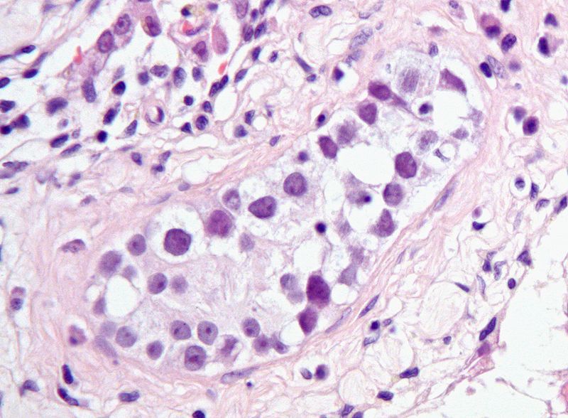

Distributed by Wikimedia
under a Creative Commons License
The germ cell precancer, itgcn, is a collection of atypical gonocytic cells lining seminiferous tubules in the testis.

_nodal_metastasis.jpg)
Distributed by Wikimedia
under a GNU License
Seminoma cells closely resemble the cells of itgcn, from which they derive (with the rare exception of the so-called spermatocytic seminoma, which behaves unlike the other types of seminomas).
The same precancer (itgcn) precedes the development of most of the invasive non-germinomatous germ cell tumors of the testis.
So, the terminologic mystery is solved. The germinomatous and the non-germinomatous germ cell tumors are classified together because most of them are derived from neoplastic intratubular germ cells (i.e., intratubular germ cell neoplasia).
But solving the terminologic mystery does not help us understand the biology of what's happening. Why does itgcn give rise to tumors of germ cells (e.g., seminomas) and to tumors of primimitive non-germ cells (e.g., embryonal carcinoma, choriocarcinoma)? How can a tumor be derived from cells that have a committed lineage (i.e., sperm cells in the case of males) that is completely unrelated to the lineages found in the tumor?
There's an answer. It has a lot to do with a phenomenon unique to germ cells called epigenomic erasure. This will be the topic of the next blog in our series on germ cell tumors.
Jump to Tomorrow's Blog
- © 2010 Jules Berman
key words: carcinogenesis, neoplasia, neoplasms, tumor development, tumour development, germ cell tumor, germ cell tumour, tumor epidemiology, increasing germ cell cancer rates, germ cell cancer, seminomas, seminomatous, non-seminomatous, non-germinomatous, embryonal carcinoma, choriocarcinoma, testis, testes, itgcn, intratubular germ cell neoplasm
In June, 2014, my book, entitled Rare Diseases and Orphan Drugs: Keys to Understanding and Treating the Common Diseases was published by Elsevier. The book builds the argument that our best chance of curing the common diseases will come from studying and curing the rare diseases.
I urge you to read more about my book. There's a generous preview of the book at the Google Books site. If you like the book, please request your librarian to purchase a copy of this book for your library or reading room.
Saturday, October 2, 2010
Germ cell tumors: definition problems
OK, getting back to the prior post on germ cell tumors, we found that the rate of occurrence of seminomatous germ cell tumors of the testes has been greatly increasing, in the white male population, since (at least) 1973. During the same period, the rate of occurrence of the other type of germ cell tumors (non-seminomatous) has hardly increased at all, for white men.
Why has the rate of occurrence of seminomas increased since 1973, in the white male population? Also, if seminomatous and non-seminomatous germ cell tumors are just morphologic variants of the same basic tumor (i.e., germ cell tumor), why wouldn't they both increase to the same extent?
Perhaps some of the problem relates to the definition of these two tumors.
Seminomas are tumors of gonocytes, a differentiated cell committed to producing gametes (sperm in males, eggs in females), or a committed progenitor cell of gamete-producing cells (i.e., an ancestral cell of a gamete-producing cell). Since seminomas are considered the neoplastic equivalent of gonocytes, there seems to be little leeway in their classification: they must be included among the germ cell tumors.
But what about the other type of germ cell tumors. This other type is known by two different names that tell us a lot about the ambivalent nature of the tumor:
From wikipedia:
"The nongerminomatous or nonseminomatous germ cell tumors (NGGCT, NSGCT) include all other germ cell tumors, pure and mixed."
How can a germ cell tumor be non-germinomatous? Wouldn't the adjective "non-germinomatous" pretty much tell you that the tumor can't be a germ cell tumor?
It reminds me of one of my favorite limericks.
As I was sitting in my chair,
I sensed the bottom was not there.
Nor legs, nor back,
But I just sat,
Ignoring little things like that.
- Anonymous
Well, what are the non-germinomatous germ cell tumors? These are tumors that usually arise in the gonads and are composed of primitive pluripotent cells. We can find pure or mixed populations of embryonal carcinoma, teratomatous tissue, and choriocarcinoma in the non-germinomatous germ cell tumors. These are the same cells that are found in the very earliest embryo and placenta. But these primitive cell types are not gonocytes (i.e., they are not differentiated cells committed to producing sperm or eggs). These tumors are composed of primitive non-germ cells.
So why are the primitive non-germ cell tumors included among the germ cell tumors?
The answer to this question comes from our understanding of the common precancer of most of the seminomatous and non-seminomatous germ cell tumors: intratubular germ cell neoplasia.
In the next several blogs, we'll discuss germ cell precancer, and we'll explain the unifying concept of germ cell neoplasia. We'll also see how a study of precancers is crucial to our understanding of neoplasia, in general.
Jump to Tomorrow's Blog
- © 2010 Jules Berman
key words: carcinogenesis, neoplasia, neoplasms, tumor development, tumour development, germ cell tumor, germ cell tumour, tumor epidemiology, increasing germ cell cancer rates, germ cell cancer, seminomas, seminomatous
In June, 2014, my book, entitled Rare Diseases and Orphan Drugs: Keys to Understanding and Treating the Common Diseases was published by Elsevier. The book builds the argument that our best chance of curing the common diseases will come from studying and curing the rare diseases.
I urge you to read more about my book. There's a generous preview of the book at the Google Books site. If you like the book, please request your librarian to purchase a copy of this book for your library or reading room.
Why has the rate of occurrence of seminomas increased since 1973, in the white male population? Also, if seminomatous and non-seminomatous germ cell tumors are just morphologic variants of the same basic tumor (i.e., germ cell tumor), why wouldn't they both increase to the same extent?
Perhaps some of the problem relates to the definition of these two tumors.
Seminomas are tumors of gonocytes, a differentiated cell committed to producing gametes (sperm in males, eggs in females), or a committed progenitor cell of gamete-producing cells (i.e., an ancestral cell of a gamete-producing cell). Since seminomas are considered the neoplastic equivalent of gonocytes, there seems to be little leeway in their classification: they must be included among the germ cell tumors.
But what about the other type of germ cell tumors. This other type is known by two different names that tell us a lot about the ambivalent nature of the tumor:
From wikipedia:
"The nongerminomatous or nonseminomatous germ cell tumors (NGGCT, NSGCT) include all other germ cell tumors, pure and mixed."
How can a germ cell tumor be non-germinomatous? Wouldn't the adjective "non-germinomatous" pretty much tell you that the tumor can't be a germ cell tumor?
It reminds me of one of my favorite limericks.
As I was sitting in my chair,
I sensed the bottom was not there.
Nor legs, nor back,
But I just sat,
Ignoring little things like that.
- Anonymous
Well, what are the non-germinomatous germ cell tumors? These are tumors that usually arise in the gonads and are composed of primitive pluripotent cells. We can find pure or mixed populations of embryonal carcinoma, teratomatous tissue, and choriocarcinoma in the non-germinomatous germ cell tumors. These are the same cells that are found in the very earliest embryo and placenta. But these primitive cell types are not gonocytes (i.e., they are not differentiated cells committed to producing sperm or eggs). These tumors are composed of primitive non-germ cells.
So why are the primitive non-germ cell tumors included among the germ cell tumors?
The answer to this question comes from our understanding of the common precancer of most of the seminomatous and non-seminomatous germ cell tumors: intratubular germ cell neoplasia.
In the next several blogs, we'll discuss germ cell precancer, and we'll explain the unifying concept of germ cell neoplasia. We'll also see how a study of precancers is crucial to our understanding of neoplasia, in general.
Jump to Tomorrow's Blog
- © 2010 Jules Berman
key words: carcinogenesis, neoplasia, neoplasms, tumor development, tumour development, germ cell tumor, germ cell tumour, tumor epidemiology, increasing germ cell cancer rates, germ cell cancer, seminomas, seminomatous
In June, 2014, my book, entitled Rare Diseases and Orphan Drugs: Keys to Understanding and Treating the Common Diseases was published by Elsevier. The book builds the argument that our best chance of curing the common diseases will come from studying and curing the rare diseases.
I urge you to read more about my book. There's a generous preview of the book at the Google Books site. If you like the book, please request your librarian to purchase a copy of this book for your library or reading room.
Friday, October 1, 2010
Povray script for book cover
Currently, I'm writing a series of blogs on germ cell tumors. But I took a break today to prepare a web page that provides the PovRay script for the cover image of my new book, Methods in Medical Informatics, Fundamental of Healthcare Programming in Perl, Python, and Ruby.

The PovRay script and a high-resoluion version of the image used in the book cover, are available at:
http://www.julesberman.info/book/coverhow.htm
I'll resume the germ cell tumor series soon.
- Jules Berman
Science is not a collection of facts. Science is what facts teach us; what we can learn about our universe, and ourselves, by deductive thinking. From observations of the night sky, made without the aid of telescopes, we can deduce that the universe is expanding, that the universe is not infinitely old, and why black holes exist. Without resorting to experimentation or mathematical analysis, we can deduce that gravity is a curvature in space-time, that the particles that compose light have no mass, that there is a theoretical limit to the number of different elements in the universe, and that the earth is billions of years old. Likewise, simple observations on animals tell us much about the migration of continents, the evolutionary relationships among classes of animals, why the nuclei of cells contain our genetic material, why certain animals are long-lived, why the gestation period of humans is 9 months, and why some diseases are rare and other diseases are common. In “Armchair Science”, the reader is confronted with 129 scientific mysteries, in cosmology, particle physics, chemistry, biology, and medicine. Beginning with simple observations, step-by-step analyses guide the reader toward solutions that are sometimes startling, and always entertaining. “Armchair Science” is written for general readers who are curious about science, and who want to sharpen their deductive skills.

The PovRay script and a high-resoluion version of the image used in the book cover, are available at:
http://www.julesberman.info/book/coverhow.htm
I'll resume the germ cell tumor series soon.
- Jules Berman
Science is not a collection of facts. Science is what facts teach us; what we can learn about our universe, and ourselves, by deductive thinking. From observations of the night sky, made without the aid of telescopes, we can deduce that the universe is expanding, that the universe is not infinitely old, and why black holes exist. Without resorting to experimentation or mathematical analysis, we can deduce that gravity is a curvature in space-time, that the particles that compose light have no mass, that there is a theoretical limit to the number of different elements in the universe, and that the earth is billions of years old. Likewise, simple observations on animals tell us much about the migration of continents, the evolutionary relationships among classes of animals, why the nuclei of cells contain our genetic material, why certain animals are long-lived, why the gestation period of humans is 9 months, and why some diseases are rare and other diseases are common. In “Armchair Science”, the reader is confronted with 129 scientific mysteries, in cosmology, particle physics, chemistry, biology, and medicine. Beginning with simple observations, step-by-step analyses guide the reader toward solutions that are sometimes startling, and always entertaining. “Armchair Science” is written for general readers who are curious about science, and who want to sharpen their deductive skills.
Monday, September 27, 2010
Non-seminomatous germ cell tumors
In yesterday's blog, we examined the enormous increase in the incidence of seminomatous germ cell tumors occurring in white non-Hispanic males.
There are two categories of germ cell tumors: seminomatous and non-seminomatous.
The seminomatous tumors are tumors composed predominantly of a single cell type, the gonocyte. The non-neoplastic gonocyte would normally produce sperm cell in the testis. Seminomas are permitted to contain a few neoplastic trophoblasts, but otherwise, seminomas are composed of a population of large, round, monomorphic cells.
The other type of germ cell tumors is the non-seminomatous tumors, and these tumors are composed of malignant cells resembling those of the pluripotent primitive embryonic (from the early embryo) or extra-embryonic (from the placenta) malignant cells. Consequently, the non-seminomatous germ cell tumors may be teratomatous, primative embryonic, choriocarcinomatous, or some mixture of these. Non-seminomatous germ cell tumors may even contain foci of seminoma! The key characteristic of non-seminomatous germ cell tumors of the testis is that they must have a component of primitive neoplastic cells that are not seminoma cells.
Can we observe the same increased incidence of non-seminomatous germ cell tumors as we saw (yesterday) in the seminomatous germ cell tumors.
NO. Here are the numbers, computed from the SEER (the U.S. National Cancer Institute's Surveillance Epidemiology and End Results) public use data files. The first column is the crude number of occurrences of non-seminomatous germ cell tumors of testes in white, non-Hispanic males. The second column is the number of occurrences expressed as a proportion of all of the seer cases for the year examined, and the third column is the number of occurrences expressed as a proportion of the U.S. population for the year examined.
Here's the graph. The blue columns are the crude occurrences. The maroon columns are the numbers as a proportion of the year's seer records, and the white column are the numbers as a porportion of the U.S. population in the examined year.

There's a small increase since 1973, but much of the increase is accounted for by the increase in the SEER population and the increase in the U.S. population for the same years. The relative (population adjusted) rate of occurrence of non-seminomatous germ cell tumors has not increased by much; certainly nothing like the increase seen yesterday, for the seminomatous germ cell tumors.
What about the germ cell tumors that occur outside the gonads? Are they increasing in occurrence since 1973? Though germ cell tumors can occur outside the gonads, they are very rare. Here are the SEER numbers for non-seminomatous non-testicular germ cell tumors in white non-Hispanic males.
Here are the numbers for seminomatous non-testicular germ cell tumors in white non-Hispanic males.
Non-testicular germ cell tumors represent a tiny fraction of the germ cell tumors occurring in men. For the purposes of analysis, there's not much you can do with these tumors. They're not going to give you statistically significant results when you try to test a hypothesis; some of them may represent misdiagnoses (e.g., colon cancer mistaken for monomorphic teratoma in a peri-testicular appendage), or a conservative topographic assignment (peri-testicular metastasis from a regressed primary germ cell tumor).
So, for the purposes of this blog, where we're trying to find a biological explanation for the rise in seminomas in non-Hispanic white males, we'll ignore the non-testicular germ cell tumors.
Jump to Tomorrow's Blog
- © 2010 Jules Berman
key words: carcinogenesis, neoplasia, neoplasms, tumor development, tumour development, germ cell tumor, germ cell tumour, tumor epidemiology, increasing germ cell cancer rates, germ cell cancer, seminomas, seminomatous, common disease, orphan disease, orphan drugs, genetics of disease, disease genetics, rules of disease biology, rare disease, pathology
In June, 2014, my book, entitled Rare Diseases and Orphan Drugs: Keys to Understanding and Treating the Common Diseases was published by Elsevier. The book builds the argument that our best chance of curing the common diseases will come from studying and curing the rare diseases.
I urge you to read more about my book. There's a generous preview of the book at the Google Books site.
There are two categories of germ cell tumors: seminomatous and non-seminomatous.
The seminomatous tumors are tumors composed predominantly of a single cell type, the gonocyte. The non-neoplastic gonocyte would normally produce sperm cell in the testis. Seminomas are permitted to contain a few neoplastic trophoblasts, but otherwise, seminomas are composed of a population of large, round, monomorphic cells.
The other type of germ cell tumors is the non-seminomatous tumors, and these tumors are composed of malignant cells resembling those of the pluripotent primitive embryonic (from the early embryo) or extra-embryonic (from the placenta) malignant cells. Consequently, the non-seminomatous germ cell tumors may be teratomatous, primative embryonic, choriocarcinomatous, or some mixture of these. Non-seminomatous germ cell tumors may even contain foci of seminoma! The key characteristic of non-seminomatous germ cell tumors of the testis is that they must have a component of primitive neoplastic cells that are not seminoma cells.
Can we observe the same increased incidence of non-seminomatous germ cell tumors as we saw (yesterday) in the seminomatous germ cell tumors.
NO. Here are the numbers, computed from the SEER (the U.S. National Cancer Institute's Surveillance Epidemiology and End Results) public use data files. The first column is the crude number of occurrences of non-seminomatous germ cell tumors of testes in white, non-Hispanic males. The second column is the number of occurrences expressed as a proportion of all of the seer cases for the year examined, and the third column is the number of occurrences expressed as a proportion of the U.S. population for the year examined.
       crude   of SEER  of U.S. Pop
1973   000109   000196   000051
1974   000147   000218   000068
1975   000157   000213   000072
1976   000165   000218   000075
1977   000189   000246   000085
1978   000167   000214   000075
1979   000182   000226   000080
1980   000216   000260   000095
1981   000222   000259   000096
1982   000203   000234   000087
1983   000226   000251   000096
1984   000219   000234   000092
1985   000238   000243   000100
1986   000233   000232   000097
1987   000253   000238   000104
1988   000222   000206   000090
1989   000263   000238   000106
1990   000243   000209   000097
1991   000237   000192   000094
1992   000237   000185   000092
1993   000245   000194   000095
1994   000222   000176   000085
1995   000216   000169   000082
1996   000247   000202   000093
1997   000225   000178   000084
1998   000234   000180   000086
1999   000245   000185   000089
2000   000237   000177   000084
2001   000223   000162   000078
2002   000258   000185   000089
2003   000230   000166   000079
2004   000278   000192   000094
2005   000275   000188   000092
2006   000251   000169   000084
2007   000277   000182   000091
Here's the graph. The blue columns are the crude occurrences. The maroon columns are the numbers as a proportion of the year's seer records, and the white column are the numbers as a porportion of the U.S. population in the examined year.

There's a small increase since 1973, but much of the increase is accounted for by the increase in the SEER population and the increase in the U.S. population for the same years. The relative (population adjusted) rate of occurrence of non-seminomatous germ cell tumors has not increased by much; certainly nothing like the increase seen yesterday, for the seminomatous germ cell tumors.
What about the germ cell tumors that occur outside the gonads? Are they increasing in occurrence since 1973? Though germ cell tumors can occur outside the gonads, they are very rare. Here are the SEER numbers for non-seminomatous non-testicular germ cell tumors in white non-Hispanic males.
       crude   of SEER  of U.S. Pop
1973   000011   000019   000005
1974   000008   000011   000003
1975   000014   000019   000006
1976   000011   000014   000005
1977   000011   000014   000004
1978   000011   000014   000004
1979   000019   000023   000008
1980   000021   000025   000009
1981   000016   000018   000006
1982   000019   000021   000008
1983   000018   000020   000007
1984   000012   000012   000005
1985   000019   000019   000007
1986   000014   000013   000005
1987   000025   000023   000010
1988   000016   000014   000006
1989   000020   000018   000008
1990   000011   000009   000004
1991   000023   000018   000009
1992   000011   000008   000004
1993   000018   000014   000006
1994   000012   000009   000004
1995   000010   000007   000003
1996   000009   000007   000003
1997   000018   000014   000006
1998   000010   000007   000003
1999   000013   000009   000004
2000   000005   000003   000001
2001   000015   000010   000005
2002   000013   000009   000004
2003   000010   000007   000003
2004   000017   000011   000005
2005   000020   000013   000006
2006   000011   000007   000003
2007   000012   000007   000003
Here are the numbers for seminomatous non-testicular germ cell tumors in white non-Hispanic males.
       crude   of SEER  of U.S. Pop
1973   000002   000003   000000
1974   000001   000001   000000
1975   000005   000006   000002
1976   000004   000005   000001
1977   000010   000013   000004
1978   000008   000010   000003
1979   000004   000004   000001
1980   000014   000016   000006
1981   000016   000018   000006
1982   000011   000012   000004
1983   000011   000012   000004
1984   000011   000011   000004
1985   000013   000013   000005
1986   000016   000015   000006
1987   000014   000013   000005
1988   000011   000010   000004
1989   000016   000014   000006
1990   000010   000008   000004
1991   000011   000008   000004
1992   000014   000010   000005
1993   000013   000010   000005
1994   000023   000018   000008
1995   000017   000013   000006
1996   000020   000016   000007
1997   000018   000014   000006
1998   000010   000007   000003
1999   000009   000006   000003
2000   000016   000012   000005
2001   000014   000010   000004
2002   000017   000012   000005
2003   000016   000011   000005
2004   000022   000015   000007
2005   000026   000017   000008
2006   000014   000009   000004
2007   000017   000011   000005
Non-testicular germ cell tumors represent a tiny fraction of the germ cell tumors occurring in men. For the purposes of analysis, there's not much you can do with these tumors. They're not going to give you statistically significant results when you try to test a hypothesis; some of them may represent misdiagnoses (e.g., colon cancer mistaken for monomorphic teratoma in a peri-testicular appendage), or a conservative topographic assignment (peri-testicular metastasis from a regressed primary germ cell tumor).
So, for the purposes of this blog, where we're trying to find a biological explanation for the rise in seminomas in non-Hispanic white males, we'll ignore the non-testicular germ cell tumors.
Jump to Tomorrow's Blog
- © 2010 Jules Berman
key words: carcinogenesis, neoplasia, neoplasms, tumor development, tumour development, germ cell tumor, germ cell tumour, tumor epidemiology, increasing germ cell cancer rates, germ cell cancer, seminomas, seminomatous, common disease, orphan disease, orphan drugs, genetics of disease, disease genetics, rules of disease biology, rare disease, pathology
In June, 2014, my book, entitled Rare Diseases and Orphan Drugs: Keys to Understanding and Treating the Common Diseases was published by Elsevier. The book builds the argument that our best chance of curing the common diseases will come from studying and curing the rare diseases.
I urge you to read more about my book. There's a generous preview of the book at the Google Books site.
Germ cell tumors: the problems
Germ cell tumors are very rare neoplasms that occur most often in young adults and children.
For a variety of reasons, which I'll try to explain in the next few blog posts, much of what we think we understand about these tumors is highly confusing and probably wrong.
Considering that these are rare tumors, you might accept a certain degree of ignorance, but sometimes the mysteries that surround rare tumors must be solved before we can make any headway understanding the more common tumors.
Also, for some strange reason, the incidence of seminomatous germ cell tumors of the testes, in the white population, has been increasing over the past 35 years (at least).
Here are the numbers, computed from the SEER (the U.S. National Cancer Institute's Surveillance Epidemiology and End Results) public use data files. The first column is the crude number of occurrences of seminomatous germ cell tumors of testes in white, non-hispanic males. The second column is the number of occurrences expressed as a proportion of all of the seer cases for the year examined, and the third column is the number of occurrences expressed as a proportion of the U.S. population for the year examined.
Here's the graph. The blue columns are the crude numbers. The maroon columns are the numbers as a proportion of the year's seer records, and the white column are the numbers as a porportion of the U.S. population in the examined year.

When the incidence of a tumor increases almost every year, and we're clueless to explain the increase, it's probably worth thinking about the problem.
Jump to Tomorrow's Blog
- © 2010 Jules Berman
key words: carcinogenesis, neoplasia, neoplasms, tumor development, tumour development, germ cell tumor, germ cell tumour, tumor epidemiology, increasing germ cell cancer rates, germ cell cancer, seminomas, seminomatous, common disease, orphan disease, orphan drugs, genetics of disease, disease genetics, rules of disease biology, rare disease, pathology
In June, 2014, my book, entitled Rare Diseases and Orphan Drugs: Keys to Understanding and Treating the Common Diseases was published by Elsevier. The book builds the argument that our best chance of curing the common diseases will come from studying and curing the rare diseases.
I urge you to read more about my book. There's a generous preview of the book at the Google Books site.
For a variety of reasons, which I'll try to explain in the next few blog posts, much of what we think we understand about these tumors is highly confusing and probably wrong.
Considering that these are rare tumors, you might accept a certain degree of ignorance, but sometimes the mysteries that surround rare tumors must be solved before we can make any headway understanding the more common tumors.
Also, for some strange reason, the incidence of seminomatous germ cell tumors of the testes, in the white population, has been increasing over the past 35 years (at least).
Here are the numbers, computed from the SEER (the U.S. National Cancer Institute's Surveillance Epidemiology and End Results) public use data files. The first column is the crude number of occurrences of seminomatous germ cell tumors of testes in white, non-hispanic males. The second column is the number of occurrences expressed as a proportion of all of the seer cases for the year examined, and the third column is the number of occurrences expressed as a proportion of the U.S. population for the year examined.
       crude   of SEER  of U.S. Pop
1973   000036   000064   000016
1974   000026   000038   000012
1975   000044   000059   000020
1976   000069   000091   000031
1977   000197   000257   000089
1978   000169   000216   000075
1979   000192   000239   000085
1980   000225   000271   000099
1981   000200   000234   000087
1982   000240   000277   000103
1983   000257   000286   000109
1984   000252   000270   000106
1985   000256   000262   000107
1986   000293   000292   000122
1987   000302   000285   000124
1988   000299   000278   000122
1989   000343   000311   000138
1990   000338   000290   000135
1991   000303   000245   000120
1992   000352   000274   000138
1993   000340   000269   000131
1994   000385   000305   000147
1995   000303   000237   000115
1996   000371   000304   000139
1997   000379   000300   000141
1998   000408   000315   000150
1999   000363   000274   000133
2000   000413   000310   000146
2001   000409   000297   000143
2002   000398   000285   000138
2003   000371   000268   000127
2004   000400   000277   000136
2005   000382   000262   000129
2006   000374   000252   000125
2007   000378   000249   000125
Here's the graph. The blue columns are the crude numbers. The maroon columns are the numbers as a proportion of the year's seer records, and the white column are the numbers as a porportion of the U.S. population in the examined year.

When the incidence of a tumor increases almost every year, and we're clueless to explain the increase, it's probably worth thinking about the problem.
Jump to Tomorrow's Blog
- © 2010 Jules Berman
key words: carcinogenesis, neoplasia, neoplasms, tumor development, tumour development, germ cell tumor, germ cell tumour, tumor epidemiology, increasing germ cell cancer rates, germ cell cancer, seminomas, seminomatous, common disease, orphan disease, orphan drugs, genetics of disease, disease genetics, rules of disease biology, rare disease, pathology
In June, 2014, my book, entitled Rare Diseases and Orphan Drugs: Keys to Understanding and Treating the Common Diseases was published by Elsevier. The book builds the argument that our best chance of curing the common diseases will come from studying and curing the rare diseases.
I urge you to read more about my book. There's a generous preview of the book at the Google Books site.
Sunday, September 26, 2010
Tumor speciation revisited
"Things that are new are wont to be set forward rudely and formlessly, and then must be polished and perfected in succeeding centuries."
- Pappus, a Helenistic mathematician (circa 350-300 B.C.E.)
Regular readers of this blog all know that I have a keen interest in the tumor speciation (why we encounter a the set of tumor types that are familiar to all pathologists, and no others). Some examples of different types of tumors are: follicular lymphomas, glioblastomas, oligodendrogliomas, seminomas, hepatocellular carcinomas, etc.)
I find the question of tumor speciation to be profound for the following reason: Research in the genetics of tumors has found that tumors are incredibly complex, with some tumors having thousands of genetic mutations, making each tumor unique from every other tumor that has ever occurred in humans. If every tumor is unique, and if many tumors are genetically complex (many mutations) and internally heterogeneous (a tumor cell may be genetically separable from another tumor cell from the same tumor), then why are there only a finite number of different kinds of tumors? Shouldn't there be a near-infinite number of tumor types?
In a prior post, I tried to answer this question, drawing an analogy from animal speciation, and though the argument seems valid, I can see how it might confuse readers. I spent much of a chapter in my Neoplasms book explaining tumor speciation, but I can't help but wonder if there's a shorter explanation.
Here's my third try:
Basically, cancer is caused by alterations in the genome. Tumor speciation is restricted to a relatively small set of patterns in the epigenome.
That's it! Here's the explanation of what it means and why it makes sense.
When oncogenic mutations occur in cells, a malignant phenotype can only arise in cells that have a specific type of differentiation. The type of differentiation that a cell manifests is determined by the epigenome (the non-sequence modifications to DNA). There are about 200 different cell types in the body. Each cell type within an individual animal has the same exact genome (DNA sequence) as every other cell type in the same animal. Neutrophils, enterocytes, neurons, thyroid cells differ from one another because of differences in their epigenomes.
Because we only observe about 200 different cell types in the body, it's likely that only a finite set of epigenomic patterns "work"; i.e., sustain cell viability. In the case of cancer, the cancer genotype can only manifest itself within a finite set of epigenomic patterns. Specific types of genetic alterations are found in specific types of epigenomic patterns (e.g., the bcr/abl mutation is seen in myeloid lineage cells). Even when cancer mutations become complex, their malignant phenotype can only occur in a restricted epigenomic background.
What happens during the process of carcinogenesis (the period following a carcinogenic mutation and leading to the emergence of an invasive cancer, often years later)? Maybe carcinogenesis requires epigenomic accommodation of the cancer genotype. Over multiple cell generations, the epigenome continuously changes until a stable epigenomic pattern is selected. Because most epigenomic patterns are not viable, most cancer mutations never lead to the emergence of a tumor.
This argument hardly constitutes proof of anything, but in my Neoplasms book, I provide many examples of how tumors develop within the constraints of allowable epigenomic patterns (i.e., the observed differentiated cell types).
Jump to the next Specified Life blog
- © 2010 Jules Berman
In June, 2014, my book, entitled Rare Diseases and Orphan Drugs: Keys to Understanding and Treating the Common Diseases was published by Elsevier. The book builds the argument that our best chance of curing the common diseases will come from studying and curing the rare diseases.
I urge you to read more about my book. There's a generous preview of the book at the Google Books site.
tags: biology of rare diseases, common diseases, genetic disease, disease genetics, orphan diseases, orphan drugs, rare disease organizations, rare disease research, rare diseases,rare disease funding, rare disease research, funding for rare diseases, importance of rare diseases, funding opportunities, books about rare diseases, books about orphan drugs, orphan drug development, pathology of rare diseases, complex diseases, carcinogenesis, neoplasia, neoplasms, oncogenes, cancer development, epigenome, genome, cancer diversity, cancer phenotype, cancer genotype, cancer epigenotype, tumor development, tumour development
- Pappus, a Helenistic mathematician (circa 350-300 B.C.E.)
Regular readers of this blog all know that I have a keen interest in the tumor speciation (why we encounter a the set of tumor types that are familiar to all pathologists, and no others). Some examples of different types of tumors are: follicular lymphomas, glioblastomas, oligodendrogliomas, seminomas, hepatocellular carcinomas, etc.)
I find the question of tumor speciation to be profound for the following reason: Research in the genetics of tumors has found that tumors are incredibly complex, with some tumors having thousands of genetic mutations, making each tumor unique from every other tumor that has ever occurred in humans. If every tumor is unique, and if many tumors are genetically complex (many mutations) and internally heterogeneous (a tumor cell may be genetically separable from another tumor cell from the same tumor), then why are there only a finite number of different kinds of tumors? Shouldn't there be a near-infinite number of tumor types?
In a prior post, I tried to answer this question, drawing an analogy from animal speciation, and though the argument seems valid, I can see how it might confuse readers. I spent much of a chapter in my Neoplasms book explaining tumor speciation, but I can't help but wonder if there's a shorter explanation.
Here's my third try:
Basically, cancer is caused by alterations in the genome. Tumor speciation is restricted to a relatively small set of patterns in the epigenome.
That's it! Here's the explanation of what it means and why it makes sense.
When oncogenic mutations occur in cells, a malignant phenotype can only arise in cells that have a specific type of differentiation. The type of differentiation that a cell manifests is determined by the epigenome (the non-sequence modifications to DNA). There are about 200 different cell types in the body. Each cell type within an individual animal has the same exact genome (DNA sequence) as every other cell type in the same animal. Neutrophils, enterocytes, neurons, thyroid cells differ from one another because of differences in their epigenomes.
Because we only observe about 200 different cell types in the body, it's likely that only a finite set of epigenomic patterns "work"; i.e., sustain cell viability. In the case of cancer, the cancer genotype can only manifest itself within a finite set of epigenomic patterns. Specific types of genetic alterations are found in specific types of epigenomic patterns (e.g., the bcr/abl mutation is seen in myeloid lineage cells). Even when cancer mutations become complex, their malignant phenotype can only occur in a restricted epigenomic background.
What happens during the process of carcinogenesis (the period following a carcinogenic mutation and leading to the emergence of an invasive cancer, often years later)? Maybe carcinogenesis requires epigenomic accommodation of the cancer genotype. Over multiple cell generations, the epigenome continuously changes until a stable epigenomic pattern is selected. Because most epigenomic patterns are not viable, most cancer mutations never lead to the emergence of a tumor.
This argument hardly constitutes proof of anything, but in my Neoplasms book, I provide many examples of how tumors develop within the constraints of allowable epigenomic patterns (i.e., the observed differentiated cell types).
Jump to the next Specified Life blog
- © 2010 Jules Berman
In June, 2014, my book, entitled Rare Diseases and Orphan Drugs: Keys to Understanding and Treating the Common Diseases was published by Elsevier. The book builds the argument that our best chance of curing the common diseases will come from studying and curing the rare diseases.
I urge you to read more about my book. There's a generous preview of the book at the Google Books site.
tags: biology of rare diseases, common diseases, genetic disease, disease genetics, orphan diseases, orphan drugs, rare disease organizations, rare disease research, rare diseases,
Friday, September 24, 2010
Melanoma and the precancer time machine
In an earlier post, I explained the precancer time machine phenomenon. Basically, when you successfully treat precancers, you don't see an immediate drop in the incidence of invasive cancers; you see a drop in invasive cancers at some point in the future, corresponding to the time at which the treated precancers would have developed into invasive cancers.
In the earlier post, I demonstrated that the precancer time machine seemed to apply in the case of dcis and invasive breast cancer.
In today's post, I've used the latest SEER (The U.S. National Cancer Institute's Surveillance Epidemiology and End Results) cancer data to show that there seems to be the same phenomenon going on for melanoma precancers.
The topic of treating melanoma precancers has been somewhat controversial. It would seem to be a no-brainer that we can reduce the incidence of invasive melanomas by treating melanoma precancers (dysplastic nevi and in situ melanomas). Unfortunately, there is no epidemilogiic evidence to support this assertion. Basically, the incidence of invasive melanoma seems to be rising every year, despite our best efforts to stem the tide (through the use of sunscreens, avoiding exposure to strong sunlight, and treating precancers).
The latest SEER data (which covers cancer cases from 1973 to 2007) seems to offer some hope that conditions might be improving.
Here's the graph of invasive melanome incidence in the U.S. SEER population.

The tallest bars (blue) are the crude numbers of occurrences of invasive malignant melanoma. The middle bars (maroon) are the occurrences of invasive melanoma expressed as a proportion of the total number of SEER cases. The bottom bars (white), are the occurrences of melanoma expressed as a proprotion of the total population of the U.S. in the perspective years.
Here are the numbers:
In the last two years studied (2006, 2007), the incidence of invasive malignant melanoma has dropped. Is this just a fluke, or does it indicate a real trend? There's no way to be sure, but inspection of the graph would indicate that it's the first time since 1973 when incidence has dropped two years running.
What about the corresponding incidence of in situ melanoma (the non-invasive precursor for invasive melanoma)?
Here's the graph for in situ melanoma.

Here are the numbers.
The incidence of in situ melanoma keeps going up and up. That's as it should be. When we successfully cure more and more in situ melanomas, we reduce the incidence of invasive melanomas.
The lag between the rise in incidence of the in situ lesions and the drop in incidence of the invasive lesions is due to the precancer time machine phenomenon.
Can we be sure? Not yet. Hopefully, over the next five years or so, the data will become a little more convincing.
- © 2010 Jules Berman
key words: precancer, precancerous, skin cancer, dysplastic nevi, dysplastic nevus, dysplastic naevus, dysplastic naevi, cancer mortality, cancer prevention, carcinogenesis
In June, 2014, my book, entitled Rare Diseases and Orphan Drugs: Keys to Understanding and Treating the Common Diseases was published by Elsevier. The book builds the argument that our best chance of curing the common diseases will come from studying and curing the rare diseases.
I urge you to read more about my book. There's a generous preview of the book at the Google Books site. If you like the book, please request your librarian to purchase a copy of this book for your library or reading room.
In the earlier post, I demonstrated that the precancer time machine seemed to apply in the case of dcis and invasive breast cancer.
In today's post, I've used the latest SEER (The U.S. National Cancer Institute's Surveillance Epidemiology and End Results) cancer data to show that there seems to be the same phenomenon going on for melanoma precancers.
The topic of treating melanoma precancers has been somewhat controversial. It would seem to be a no-brainer that we can reduce the incidence of invasive melanomas by treating melanoma precancers (dysplastic nevi and in situ melanomas). Unfortunately, there is no epidemilogiic evidence to support this assertion. Basically, the incidence of invasive melanoma seems to be rising every year, despite our best efforts to stem the tide (through the use of sunscreens, avoiding exposure to strong sunlight, and treating precancers).
The latest SEER data (which covers cancer cases from 1973 to 2007) seems to offer some hope that conditions might be improving.
Here's the graph of invasive melanome incidence in the U.S. SEER population.

The tallest bars (blue) are the crude numbers of occurrences of invasive malignant melanoma. The middle bars (maroon) are the occurrences of invasive melanoma expressed as a proportion of the total number of SEER cases. The bottom bars (white), are the occurrences of melanoma expressed as a proprotion of the total population of the U.S. in the perspective years.
Here are the numbers:
       crude   of SEER  of U.S. Pop
1973   001062   001916   000501
1974   001305   001938   000610
1975   001559   002116   000721
1976   001601   002116   000734
1977   001791   002337   000813
1978   001829   002347   000821
1979   001973   002460   000876
1980   002190   002645   000963
1981   002298   002690   001001
1982   002352   002714   001015
1983   002350   002617   001005
1984   002462   002638   001043
1985   002795   002864   001174
1986   002947   002942   001227
1987   003050   002879   001258
1988   002948   002743   001205
1989   003194   002896   001294
1990   003272   002816   001311
1991   003518   002853   001395
1992   003569   002786   001399
1993   003611   002860   001400
1994   003904   003097   001499
1995   004189   003283   001594
1996   004438   003641   001673
1997   004627   003668   001728
1998   004739   003663   001753
1999   004893   003704   001794
2000   005105   003832   001814
2001   005380   003911   001886
2002   005377   003857   001867
2003   005514   003993   001898
2004   005859   004062   001998
2005   006451   004431   002180
2006   006431   004335   002152
2007   006325   004172   002097
In the last two years studied (2006, 2007), the incidence of invasive malignant melanoma has dropped. Is this just a fluke, or does it indicate a real trend? There's no way to be sure, but inspection of the graph would indicate that it's the first time since 1973 when incidence has dropped two years running.
What about the corresponding incidence of in situ melanoma (the non-invasive precursor for invasive melanoma)?
Here's the graph for in situ melanoma.

Here are the numbers.
       crude   of SEER  of U.S. Pop
1973   000035   000063   000016
1974   000059   000087   000027
1975   000081   000109   000037
1976   000096   000126   000044
1977   000155   000202   000070
1978   000177   000227   000079
1979   000177   000220   000078
1980   000263   000317   000115
1981   000276   000323   000120
1982   000326   000376   000140
1983   000384   000427   000164
1984   000466   000499   000197
1985   000638   000653   000268
1986   000723   000721   000301
1987   000782   000738   000322
1988   000824   000766   000337
1989   000976   000885   000395
1990   001127   000970   000451
1991   001192   000966   000472
1992   001373   001072   000538
1993   001375   001089   000533
1994   001599   001268   000614
1995   001920   001504   000730
1996   002091   001715   000788
1997   002258   001790   000843
1998   002533   001957   000937
1999   002765   002093   001013
2000   003202   002404   001137
2001   003508   002550   001230
2002   003656   002622   001269
2003   003429   002483   001180
2004   003604   002499   001229
2005   004060   002788   001372
2006   004048   002728   001354
2007   004291   002831   001422
The incidence of in situ melanoma keeps going up and up. That's as it should be. When we successfully cure more and more in situ melanomas, we reduce the incidence of invasive melanomas.
The lag between the rise in incidence of the in situ lesions and the drop in incidence of the invasive lesions is due to the precancer time machine phenomenon.
Can we be sure? Not yet. Hopefully, over the next five years or so, the data will become a little more convincing.
- © 2010 Jules Berman
key words: precancer, precancerous, skin cancer, dysplastic nevi, dysplastic nevus, dysplastic naevus, dysplastic naevi, cancer mortality, cancer prevention, carcinogenesis
In June, 2014, my book, entitled Rare Diseases and Orphan Drugs: Keys to Understanding and Treating the Common Diseases was published by Elsevier. The book builds the argument that our best chance of curing the common diseases will come from studying and curing the rare diseases.
I urge you to read more about my book. There's a generous preview of the book at the Google Books site. If you like the book, please request your librarian to purchase a copy of this book for your library or reading room.
Wednesday, September 22, 2010
Methods in Medical Informatics
My new book, Methods in Medical Informatics: Fundamentals of Healthcare Programming in Perl, Python,and Ruby, was published yesterday by CRC Press.
I've prepared web pages with a book description and the Table of Contents for anyone interested.
- Jules Berman
Subscribe to:
Posts (Atom)