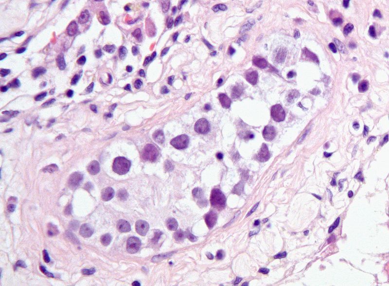
Showing posts with label precancer. Show all posts

Sunday, July 13, 2014

Treating precancers reduces breast cancer deaths

Breast cancer deaths rose through the '70s and '80s, but declined in the '90s. For nearly the past 20 years, American women have had about a 2% annual drop in the breast cancer death rate.

Here is the mortality graph provided by the National Cancer Institutes SEER (Surveillance, Epidemiology and End Results) program.


Though nobody wants to take the blame for the rise in breast cancer deaths in the '70s and '80s, lots of people want credit for the fall of breast cancer deaths that began in the '90s. Was it due to a reduction to the exposure of carcinogens, or to better treatment, or to earlier diagnosis?

The fall in breast cancer deaths does not seem to be due to cancer prevention. While the deaths from breast cancer were falling, there was an apparent rise in the incidence of breast cancer cases. Here is the SEER graph for the incidence in breast cancer in the U.S.


Since the breast cancer incidence rose while the deaths from breast cancer dropped, it seemed as though the benefit must have come from better treatment or earlier detection.

A major study, attempting to resolve this issue, was published in the New England Journal of Medicine, in 2005:

Effect of screening and adjuvant therapy on mortality from breast cancer.
Berry DA, Cronin KA, Plevritis SK, Fryback DG, Clarke L, Zelen M, Mandelblatt JS, Yakovlev AY, Habbema JD, Feuer EJ; Cancer Intervention and Surveillance Modeling Network (CISNET) Collaborators. N Engl J Med 353:1784-1792, 2005.


They concluded that that 28 to 65 percent of the sharp decrease in breast cancer
deaths from 1990 to 2000 was due to mammograms. The remainder of the improvement was was attributed improved breast cancer treatment.

The study did not take into account the great contribution of precancer treatment to the reduction of breast cancer deaths.

Let's review this SEER data, this time taking into account the diagnosis of DCIS (ductal carcinoma in situ) a precancer that precedes the development of invasive breast cancer. Here is the SEER data for the incidence of all breast cancer and of DCIS (the precancer for breast cancer).


In the past few decades, there has been a huge rise in the number of diagnosed cases of breast precancers. This is due largely to the use of mammography, which can detect lesions that cannot be found by palpation. When a precancer is detected and removed, the patient does not develop invasive cancer.

The total number of breast cancer cases includes cases of DCIS. If we subtract the number of breast precancer cases (DCIS) from the total number of breast cancer cases, we get the incidence of invasive breast cancer cases. Here is the SEER data.


The middle bars in the graph represent the incidence of invasive breast cancers. The graph shows that incidence of invasive breast cancers has actually dropped since the early '90s, as the diagnosis and treatment of DCIS has risen.

Much of the decrease in breast cancer mortality can be accounted for by the diagnosis and treatment of breast precancers. In fact the drop in breast cancer deaths follows the same slope, and has about the same magnitude, as the drop in invasive breast cancers that follows the increase in breast precancer treatments.

- © 2010 Jules Berman


Science is not a collection of facts. Science is what facts teach us; what we can learn about our universe, and ourselves, by deductive thinking. From observations of the night sky, made without the aid of telescopes, we can deduce that the universe is expanding, that the universe is not infinitely old, and why black holes exist. Without resorting to experimentation or mathematical analysis, we can deduce that gravity is a curvature in space-time, that the particles that compose light have no mass, that there is a theoretical limit to the number of different elements in the universe, and that the earth is billions of years old. Likewise, simple observations on animals tell us much about the migration of continents, the evolutionary relationships among classes of animals, why the nuclei of cells contain our genetic material, why certain animals are long-lived, why the gestation period of humans is 9 months, and why some diseases are rare and other diseases are common. In “Armchair Science”, the reader is confronted with 129 scientific mysteries, in cosmology, particle physics, chemistry, biology, and medicine. Beginning with simple observations, step-by-step analyses guide the reader toward solutions that are sometimes startling, and always entertaining. “Armchair Science” is written for general readers who are curious about science, and who want to sharpen their deductive skills.


Thursday, October 7, 2010

Germ cell cancers of testes: conclusion

This is the last entry on a series of blogs on germ cell cancers of the testes.

I've added forward and backward links for each of the blogs in the series, so you can visit the first blog in the series and click forward or backward through the sequential entries.

Basically, in this series, we showed, using the SEER public use data files, that there has been a large increase in the incidence of germ cell cancers of the testis in white non-Hispanic males since the first SEER observation year (1973) up to the most recent data year (2007).

Along with the increase in seminomatous germ cell cancers was a lesser but parallel increase in the non-seminomatous germ cell cancers of the testis, when compared in birth cohort populations.

The seminomatous and non-seminomatous germ cell cancers, though derived from very different cell types (germ cells versus embryonic/extra-embryonic primitive cells) develop from the same precanceous lesion (usually intratubular germ cell neoplasia and sometimes gonadoblastoma). Precancerous germ cells are characterized by epigenomic erasure, and this "erased" state seems to allow precancerous germ cells to develop into seminomas or into tumors derived from totipotent stem cells.

Testicular precancers develop from disorders of sex development. The incidence of disorders of sex development, like the incidence of testicular germ cell cancers, has been rising. The cause for the rise of disorders of sex development (and the concomitant rise in testicular germ cell cancers) is unknown. However, the ubiquitous appearance of the platicizer and endocrine disruptor, Bisphenol A, has captured the interest of toxicologists and cancer researchers.

All of these issues were discussed in this completed series of blogs on testicular germ cell cancers.

- © 2010 Jules Berman

key words: carcinogenesis, neoplasia, neoplasms, tumor development, tumour development, germ cell tumor, germ cell tumour, tumor epidemiology, increasing germ cell cancer rates, germ cell cancer, seminomas, seminomatous, non-seminomatous, non-germinomatous, embryonal carcinoma, choriocarcinoma, testis, testes, itgcn, intratubular germ cell neoplasm, plasticizers, endocrine disruptors

Wednesday, October 6, 2010

Explaining the rise in testicular germ cell tumors

In yesterday's blog we saw that the rise in testicular cancer rates in white males showed a parallel increase in seminomatous and non-seminomatous germ cell cancers among birth cohorts.

What can explain this increase?

There is one class of conditions that is overwhelmingly associated with the development of germ cell tumors of the testis: disorders of sex development of the testes.[1] Among the conditions within this general group are testicular dysgenesis, testicular feminization (insensitivity to androgens), and cryptorchidism. Disorders of sex development of the testis raise the incidence of intratubular germ cell neoplasia or of gonadoblastoma, both of which are testicular precancers.

As you might expect, along with the observed increase in testicular germ cell cancers in white males, there has been an observed increase in the incidence of disorders of sex development in the same population. [1,2] These disorders are characterized by a retardation in the maturation of primordial germ cells, along with an apparent mitotic over-stimulation of these same cells: leading to a proliferative, precancerous condition.

Though there is no proof at the moment, we might expect that males who develop testicular germ cell cancer who have clinically normal testes, may harbor small foci of [clinically unobserved] germ cell proliferative lesions.

What has caused the increased incidence of disorders of sex development in the testes? We don't know, but we have a candidate: the ubiquitous plasticizer and endocrine disruptor, Bisphenol A.

Bisphenol A is a synthetic estrogen used in the process of manufacturing plastics, and has been detected in the serum, milk, saliva, urine, and amniotic fluid of humans.[3] Because we get our daily dose of Bisphenol A from plastic bottles, one would expect that the levels of Bisphenol A in our blood would have increased steadily over the past several decades [coinciding with our increased dependence of plastic food and drink containers]. You might also expect that if Bisphenol A produced testicular cancers, you would see the largest increases in incidence among the wealthiest populations in the most industrialized nations [as we do].

Can we assume that Bisphenol A is causing the rise of incidence of testicular germ cell cancers? Absolutely not. All of the evidence, so far, is very weak (if it can be called evidence at all!). Still, nobody would suggest that Bisphenol A has much to recommend itself as a healthy addition to our diets. It seems prudent to try to limit our exposure to this compound when feasible, particularly among infants and pregnant women.

1. Pleskacova J, Hersmus R, Oosterhuis JW, Setyawati BA, Faradz SM, Cools M, Wolffenbuttel KP, Lebl J, Drop SL, Looijenga LH. Tumor Risk in Disorders of Sex Development. Sex Dev 4:259-269, 2010.

2. Skakkebaek NE, Rajpert-De Meyts E, Jorgensen N, Main KM, Leffers H, Andersson AM, Juul A, Jensen TK, Toppari J. Testicular cancer trends as 'whistle blowers' of testicular developmental problems in populations. Int J Androl 30:198-204, 2007.

3. Bouskine A, Nebout M, Brucker-Davis F, Benahmed M, Fenichel P. Low Doses of Bisphenol A Promote Human Seminoma Cell Proliferation by Activating PKA and PKG via a Membrane G-Protein-Coupled Estrogen Receptor. Environ Health Perspect 117:1053-1058, 2009.

This series of blogs has drawn heavily from the public use SEER data sets produced by the U.S. National Cancer Institute's Surveillance, Epidemiology and End Results project. In the next blog, I'll discuss how this data can be obtained and used by the science-minded public.

Jump to Tomorrow's blog

- © 2010 Jules Berman

key words: carcinogenesis, neoplasia, neoplasms, tumor development, tumour development, germ cell tumor, germ cell tumour, tumor epidemiology, increasing germ cell cancer rates, germ cell cancer, seminomas, seminomatous, non-seminomatous, non-germinomatous, embryonal carcinoma, choriocarcinoma, testis, testes, itgcn, intratubular germ cell neoplasm, plasticizers, endocrine disruptors

About my book, Precancer: The Beginning and the End of Cancer. Nearly every type of cancer passes through a precancer phase, during which it cannot metastasize or invade other tissues. While medicine is not always successful in treating or curing advanced stages of cancers, recent advances in our understanding of carcinogenesis have helped us to develop strategies to prevent, diagnose, and treat many cancers at the precancer stage. Research in this field is escalating rapidly as the evidence increasingly shows that the number of annual cancer deaths could be drastically reduced through the effective treatment and cure of precancer lesions. This book begins by explaining why it has been so difficult to cure cancers, followed by a review of precancer biology, with descriptions of the most common precancer lesions. The final chapters provide practical socio-political and medical goals for precancer treatment, including discussions of the economics and politics of treating precancers.


I urge you to read more about this important topic. Google Books has provided a generous preview of this book.

Tuesday, October 5, 2010

Explaining the germ cell cancer rates

In yesterday's blog we explained how the precancer of testicular germ cell tumors, intratubular germ cell neoplasia, gives rise to seminomas (differentiated germinomatous lineage) and non-seminomas (tumors of pluripotent progenitor cells that are not of germ cell lineage).

In the first blog of this series on germ cell tumors , we noted that the increase in occurrences of seminomas has outpaced the occurrences of the nonseminomatous germ cell tumors.

Here is a graph, produced from the SEER public use data sets, of the crude occurrences of seminoma and non-seminoma testicular germ cell tumors, in white males, since 1973.


The light blue bars are the seminomas, and the maroon bars are the non-seminomatous germ cell tumors of the testes. Since 1973, the seminomas increased from a number much lower than the occurrences of the non-seminomatous germ cell tumors; exceeding them in 1977. Since 1977, the crude occurrences of seminomas has greatly outpaced the occurrences of the non-seminomatous germ cell tumors of testes in white males.

Why? If both types of tumors are coming from the same precancer, why are their trends of occurrence non-parallel?

Well, there are several possible answers. It is possible that some external influence has modified the step in the progression of precancer to cancer, to favor the occurrence of seminomas.

However, it is also possible that their increases in occurrence are indeed parallel, and we're just not seeing it in our graph. Bray et al have looked at the incidence of testicular seminoma and non-seminoma germ cell tumors, by cohort (i.e., year of birth), not by year of occurrence.[1]

Bray F, Richiardi L, Ekbom A, Forman D,Pukkala E, Cuninkova M, Moller H. Do Testicular Seminoma and Nonseminoma Share the Same Etiology? Evidence from an Age-Period-Cohort Analysis of Incidence Trends in Eight European Countries. Cancer Epidemiol Biomarkers Prev 15:652–658, 2006.

When the comparisons are based on cohort (comparing incidence for people born the same year), most of the differences vanish [between the incidence of seminomas and non-seminomatous germ cell tumors].

When do we see a birth corhort effect on tumor incidence? For cancers, a cohort effect is best observed when individuals born in one year are exposed (as a population group) to a causal agent that is different from the exposure of individuals born in other years. The cancers that result may occur at many different ages, thus erasing the cohort effect when the data is stratified by year of occurrence (as we had done the graph above). Only when you look at the birth cohort will you find a trend that may relate to a carcinogenic exposure.

OK. The birth cohort data reported by Bray et al would seem to indicate that some generational effect is acting on succeeding cohorts to produce a shared increase in the incidence of all testicular germ cell cancers. Furthermore, whatever is causing the generational effect is likely to be of short duration or differ significantly from year to year. Why is that? If the exposure of a carcinogen were of long duration or were the same from year to year, then every cohort would be exposed similarly, and there would be no birth-year specific effect.

So, now the mystery is: What are the conditions and carcinogens that might cause testicular germ cell tumors, which have changed, year-by-year, to produce the observed rise in these cancers in white males? This will be the topic of the next blog.

Jump to Tomorrow's Blog

- © 2010 Jules Berman

key words: carcinogenesis, neoplasia, neoplasms, tumor development, tumour development, germ cell tumor, germ cell tumour, tumor epidemiology, increasing germ cell cancer rates, germ cell cancer, seminomas, seminomatous, non-seminomatous, non-germinomatous, embryonal carcinoma, choriocarcinoma, testis, testes, itgcn, intratubular germ cell neoplasm
Science is not a collection of facts. Science is what facts teach us; what we can learn about our universe, and ourselves, by deductive thinking. From observations of the night sky, made without the aid of telescopes, we can deduce that the universe is expanding, that the universe is not infinitely old, and why black holes exist. Without resorting to experimentation or mathematical analysis, we can deduce that gravity is a curvature in space-time, that the particles that compose light have no mass, that there is a theoretical limit to the number of different elements in the universe, and that the earth is billions of years old. Likewise, simple observations on animals tell us much about the migration of continents, the evolutionary relationships among classes of animals, why the nuclei of cells contain our genetic material, why certain animals are long-lived, why the gestation period of humans is 9 months, and why some diseases are rare and other diseases are common. In “Armchair Science”, the reader is confronted with 129 scientific mysteries, in cosmology, particle physics, chemistry, biology, and medicine. Beginning with simple observations, step-by-step analyses guide the reader toward solutions that are sometimes startling, and always entertaining. “Armchair Science” is written for general readers who are curious about science, and who want to sharpen their deductive skills.

Sunday, October 3, 2010

Germ cell precancers and epigenetic erasure

In yesterday's blog , we left off with a question: "How it is possible for intratubular germ cell neoplasia to be the precursor for both germinomatous germ cell cancers (i.e., seminoma) and non-germinomatous germ cell cancers (e.g. embryonal carcinoma, choriocarcinoma)?

The key is epigenetic erasure, a phenomenon unique to germ cells.

As an organism develops, cells specialize into about 200 differentiated cell types. All these different types of cells have the same genetic sequence (genome). Cell types are distingued, one from the other, by epigenetic modifications. Epigenetic modifications to genes involve base methylation, conformational changes in chromosomes, protein modifications... anything other than changes in DNA sequence.

Germ cells, like all other differentiated cells, have epigenetic modifications. The unique thing about germ cells is that they must undergo epigenetic erasure prior to the production of gametes; otherwise the gametes would be imprinted with the epigenetic modifications characteristic of the parent organism and would not be capable of recombining during fertilization to produce a fully de-differentiated, totipotent product.[1]

The cells of intratubular germ cell neoplasia (the precancer of most male germ cell tumors) and of seminomas, are all characterized by DNA hypomethylation; not so for the cells of non-germinomatous germ cell tumors.[2,3] DNA Hypomethylation is seen in epigenomic erasure [of germ cells].

"Erased" germ cells are capable of developing into totipotent embryonic cells.[4] It would seem that a plausible mechanism for the development of non-germinomatous germ cell cancers from a germ cell precursor (intratubular germ cell neoplasia, itgcn) is that the "erased" itgcn cells, during cancer development, transform into totipotent cells, capable of differentiating into cells from any embryonic layer (e.g., embryonal carcinoma), or into extra-embryonic tissue (e.g., choriocarcinoma).

This explains why the itgcn, the germ cell precancer, can give rise to both germinomatous (erased) and non-germinomatous (epigenetic-modified) cancers.

There is only one mystery left to solve (the original mystery that we started with, about 4 blog entries back ). If germinomatous and non-germinomatous germ cell cancers both arise from the same precursor, why is there a much greater increase in the rate of occurrence of seminomas compared with the rate of occurrence of non-germinomatous cancers, since 1973?

1. Allegrucci C, Thurston A, Lucas E, Young L. Epigenetics and the germline. Reproduction 129:137-149, 2005.

2. Netto GJ et al.Global DNA hypomethylation in intratubular germ cell neoplasia and seminoma, but not in nonseminomatous male germ cell tumors. Modern Pathology 21: 1337-1344, 2008.

3. Lind GE, Skotheim RI, Lothe RA. The epigenome of testicular germ cell tumors. APMIS (Acta Pathologica, Microbiologica et Immunologica Scandinavica) 115:1147-1160, 2007.

4. Turnpenny L. Derivation of human embryonic germ cells: an alternative source of pluripotent stem cells. Stem Cells 21:598-609, 2003.

Jump to Tomorrow's Blog

- © 2010 Jules Berman

key words: carcinogenesis, neoplasia, neoplasms, tumor development, tumour development, germ cell tumor, germ cell tumour, tumor epidemiology, increasing germ cell cancer rates, germ cell cancer, seminomas, seminomatous, non-seminomatous, non-germinomatous, embryonal carcinoma, choriocarcinoma, testis, testes, itgcn, intratubular germ cell neoplasm


In June, 2014, my book, entitled Rare Diseases and Orphan Drugs: Keys to Understanding and Treating the Common Diseases was published by Elsevier. The book builds the argument that our best chance of curing the common diseases will come from studying and curing the rare diseases.


I urge you to read more about my book. There's a generous preview of the book at the Google Books site. If you like the book, please request your librarian to purchase a copy of this book for your library or reading room.

Germ cell tumor common precancer: itgcn

In yesterday's blog , we discussed the terminology problem with the germ cell tumors. Basically, if you have a sub-class of germ cell tumors that are called "non-germinomatous germ cell tumors," isn't that a contradiction in terms? Isn't it like saying that dehydrated water is a subclass of water?

The answer is simple: the classic germ cell tumor of the testes (seminoma), as well as most of the malignant non-germinomatous germ cell tumors of the testes, arise from the same precancer: intratubular germ cell neoplasia (itgcn). Because itgcn is composed of dysplastic (early neoplastic) germ cells, both the germinomatous and non-germinomatous tumors have a germ cell origin.

You can easily appreciate the morphologic similarity between itgcn and seminoma by looking at a histologic preparation of each.


Image of Intratubular Germ Cell neoplasia
Distributed by Wikimedia
under a Creative Commons License


The germ cell precancer, itgcn, is a collection of atypical gonocytic cells lining seminiferous tubules in the testis.


Image of Seminoma
Distributed by Wikimedia
under a GNU License


Seminoma cells closely resemble the cells of itgcn, from which they derive (with the rare exception of the so-called spermatocytic seminoma, which behaves unlike the other types of seminomas).

The same precancer (itgcn) precedes the development of most of the invasive non-germinomatous germ cell tumors of the testis.

So, the terminologic mystery is solved. The germinomatous and the non-germinomatous germ cell tumors are classified together because most of them are derived from neoplastic intratubular germ cells (i.e., intratubular germ cell neoplasia).

But solving the terminologic mystery does not help us understand the biology of what's happening. Why does itgcn give rise to tumors of germ cells (e.g., seminomas) and to tumors of primimitive non-germ cells (e.g., embryonal carcinoma, choriocarcinoma)? How can a tumor be derived from cells that have a committed lineage (i.e., sperm cells in the case of males) that is completely unrelated to the lineages found in the tumor?

There's an answer. It has a lot to do with a phenomenon unique to germ cells called epigenomic erasure. This will be the topic of the next blog in our series on germ cell tumors.

Jump to Tomorrow's Blog

- © 2010 Jules Berman

key words: carcinogenesis, neoplasia, neoplasms, tumor development, tumour development, germ cell tumor, germ cell tumour, tumor epidemiology, increasing germ cell cancer rates, germ cell cancer, seminomas, seminomatous, non-seminomatous, non-germinomatous, embryonal carcinoma, choriocarcinoma, testis, testes, itgcn, intratubular germ cell neoplasm
In June, 2014, my book, entitled Rare Diseases and Orphan Drugs: Keys to Understanding and Treating the Common Diseases was published by Elsevier. The book builds the argument that our best chance of curing the common diseases will come from studying and curing the rare diseases.


I urge you to read more about my book. There's a generous preview of the book at the Google Books site. If you like the book, please request your librarian to purchase a copy of this book for your library or reading room.

Saturday, October 2, 2010

Germ cell tumors: definition problems

OK, getting back to the prior post on germ cell tumors, we found that the rate of occurrence of seminomatous germ cell tumors of the testes has been greatly increasing, in the white male population, since (at least) 1973. During the same period, the rate of occurrence of the other type of germ cell tumors (non-seminomatous) has hardly increased at all, for white men.

Why has the rate of occurrence of seminomas increased since 1973, in the white male population? Also, if seminomatous and non-seminomatous germ cell tumors are just morphologic variants of the same basic tumor (i.e., germ cell tumor), why wouldn't they both increase to the same extent?

Perhaps some of the problem relates to the definition of these two tumors.

Seminomas are tumors of gonocytes, a differentiated cell committed to producing gametes (sperm in males, eggs in females), or a committed progenitor cell of gamete-producing cells (i.e., an ancestral cell of a gamete-producing cell). Since seminomas are considered the neoplastic equivalent of gonocytes, there seems to be little leeway in their classification: they must be included among the germ cell tumors.

But what about the other type of germ cell tumors. This other type is known by two different names that tell us a lot about the ambivalent nature of the tumor:

From wikipedia:

"The nongerminomatous or nonseminomatous germ cell tumors (NGGCT, NSGCT) include all other germ cell tumors, pure and mixed."

How can a germ cell tumor be non-germinomatous? Wouldn't the adjective "non-germinomatous" pretty much tell you that the tumor can't be a germ cell tumor?

It reminds me of one of my favorite limericks.

As I was sitting in my chair,
I sensed the bottom was not there.
Nor legs, nor back,
But I just sat,
Ignoring little things like that.


- Anonymous

Well, what are the non-germinomatous germ cell tumors? These are tumors that usually arise in the gonads and are composed of primitive pluripotent cells. We can find pure or mixed populations of embryonal carcinoma, teratomatous tissue, and choriocarcinoma in the non-germinomatous germ cell tumors. These are the same cells that are found in the very earliest embryo and placenta. But these primitive cell types are not gonocytes (i.e., they are not differentiated cells committed to producing sperm or eggs). These tumors are composed of primitive non-germ cells.

So why are the primitive non-germ cell tumors included among the germ cell tumors?

The answer to this question comes from our understanding of the common precancer of most of the seminomatous and non-seminomatous germ cell tumors: intratubular germ cell neoplasia.

In the next several blogs, we'll discuss germ cell precancer, and we'll explain the unifying concept of germ cell neoplasia. We'll also see how a study of precancers is crucial to our understanding of neoplasia, in general.

Jump to Tomorrow's Blog

- © 2010 Jules Berman

key words: carcinogenesis, neoplasia, neoplasms, tumor development, tumour development, germ cell tumor, germ cell tumour, tumor epidemiology, increasing germ cell cancer rates, germ cell cancer, seminomas, seminomatous
In June, 2014, my book, entitled Rare Diseases and Orphan Drugs: Keys to Understanding and Treating the Common Diseases was published by Elsevier. The book builds the argument that our best chance of curing the common diseases will come from studying and curing the rare diseases.


I urge you to read more about my book. There's a generous preview of the book at the Google Books site. If you like the book, please request your librarian to purchase a copy of this book for your library or reading room.

Friday, September 24, 2010

Melanoma and the precancer time machine

In an earlier post, I explained the precancer time machine phenomenon. Basically, when you successfully treat precancers, you don't see an immediate drop in the incidence of invasive cancers; you see a drop in invasive cancers at some point in the future, corresponding to the time at which the treated precancers would have developed into invasive cancers.

In the earlier post, I demonstrated that the precancer time machine seemed to apply in the case of dcis and invasive breast cancer.

In today's post, I've used the latest SEER (The U.S. National Cancer Institute's Surveillance Epidemiology and End Results) cancer data to show that there seems to be the same phenomenon going on for melanoma precancers.

The topic of treating melanoma precancers has been somewhat controversial. It would seem to be a no-brainer that we can reduce the incidence of invasive melanomas by treating melanoma precancers (dysplastic nevi and in situ melanomas). Unfortunately, there is no epidemilogiic evidence to support this assertion. Basically, the incidence of invasive melanoma seems to be rising every year, despite our best efforts to stem the tide (through the use of sunscreens, avoiding exposure to strong sunlight, and treating precancers).

The latest SEER data (which covers cancer cases from 1973 to 2007) seems to offer some hope that conditions might be improving.

Here's the graph of invasive melanome incidence in the U.S. SEER population.


The tallest bars (blue) are the crude numbers of occurrences of invasive malignant melanoma. The middle bars (maroon) are the occurrences of invasive melanoma expressed as a proportion of the total number of SEER cases. The bottom bars (white), are the occurrences of melanoma expressed as a proprotion of the total population of the U.S. in the perspective years.

Here are the numbers:

crude of SEER of U.S. Pop
1973 001062 001916 000501
1974 001305 001938 000610
1975 001559 002116 000721
1976 001601 002116 000734
1977 001791 002337 000813
1978 001829 002347 000821
1979 001973 002460 000876
1980 002190 002645 000963
1981 002298 002690 001001
1982 002352 002714 001015
1983 002350 002617 001005
1984 002462 002638 001043
1985 002795 002864 001174
1986 002947 002942 001227
1987 003050 002879 001258
1988 002948 002743 001205
1989 003194 002896 001294
1990 003272 002816 001311
1991 003518 002853 001395
1992 003569 002786 001399
1993 003611 002860 001400
1994 003904 003097 001499
1995 004189 003283 001594
1996 004438 003641 001673
1997 004627 003668 001728
1998 004739 003663 001753
1999 004893 003704 001794
2000 005105 003832 001814
2001 005380 003911 001886
2002 005377 003857 001867
2003 005514 003993 001898
2004 005859 004062 001998
2005 006451 004431 002180
2006 006431 004335 002152
2007 006325 004172 002097

In the last two years studied (2006, 2007), the incidence of invasive malignant melanoma has dropped. Is this just a fluke, or does it indicate a real trend? There's no way to be sure, but inspection of the graph would indicate that it's the first time since 1973 when incidence has dropped two years running.

What about the corresponding incidence of in situ melanoma (the non-invasive precursor for invasive melanoma)?

Here's the graph for in situ melanoma.


Here are the numbers.

crude of SEER of U.S. Pop
1973 000035 000063 000016
1974 000059 000087 000027
1975 000081 000109 000037
1976 000096 000126 000044
1977 000155 000202 000070
1978 000177 000227 000079
1979 000177 000220 000078
1980 000263 000317 000115
1981 000276 000323 000120
1982 000326 000376 000140
1983 000384 000427 000164
1984 000466 000499 000197
1985 000638 000653 000268
1986 000723 000721 000301
1987 000782 000738 000322
1988 000824 000766 000337
1989 000976 000885 000395
1990 001127 000970 000451
1991 001192 000966 000472
1992 001373 001072 000538
1993 001375 001089 000533
1994 001599 001268 000614
1995 001920 001504 000730
1996 002091 001715 000788
1997 002258 001790 000843
1998 002533 001957 000937
1999 002765 002093 001013
2000 003202 002404 001137
2001 003508 002550 001230
2002 003656 002622 001269
2003 003429 002483 001180
2004 003604 002499 001229
2005 004060 002788 001372
2006 004048 002728 001354
2007 004291 002831 001422

The incidence of in situ melanoma keeps going up and up. That's as it should be. When we successfully cure more and more in situ melanomas, we reduce the incidence of invasive melanomas.

The lag between the rise in incidence of the in situ lesions and the drop in incidence of the invasive lesions is due to the precancer time machine phenomenon.

Can we be sure? Not yet. Hopefully, over the next five years or so, the data will become a little more convincing.

- © 2010 Jules Berman

key words: precancer, precancerous, skin cancer, dysplastic nevi, dysplastic nevus, dysplastic naevus, dysplastic naevi, cancer mortality, cancer prevention, carcinogenesis
In June, 2014, my book, entitled Rare Diseases and Orphan Drugs: Keys to Understanding and Treating the Common Diseases was published by Elsevier. The book builds the argument that our best chance of curing the common diseases will come from studying and curing the rare diseases.


I urge you to read more about my book. There's a generous preview of the book at the Google Books site. If you like the book, please request your librarian to purchase a copy of this book for your library or reading room.

Saturday, September 11, 2010

Precancer time machine

In the previous post , we discussed breast precancer. We saw that as mammography picked up earlier and earlier lesions (precancers and early breast cancers), deaths from breast cancer dropped, along with the incidence of invasive breast cancer.


In this graph, which covers the years 1975 to 2007, the top line of bars (blue) represent the incidence of breast cancers (including invasive and non-invasice lesions). The next lower line of bars (maroon) is the incidence of the invasive breast cancers (the kind that account for breast cancer deaths), and the bottom line of bars represents the rate of precancers (ductal carcinoma in situ) collected by SEER.

As you recall from the previous blog , the big drop in cancer death rates did not occur until about 1990, well after there was a rise in the number of diagosed breast precancers.

Why didn't the precancers diagnosed in the 1980s produce an immediate drop in the rate of breast cancer deaths or in the incidence of invasive cancers?

Precancer treatment works like a time machine. When you cure a precancer today, you don't see a reduction in the number of cancers that would arise that same day. You see a reduction in the number of cancers that will arise in some future date (if the precancer had been allowed to develop into a cancer, over time).

If a precancer would ordinarily require 5 years to develop invasive features (i.e., become a cancer), and you diagnose and treat the precancer in 2010, then you will eliminate a cancer that would have occurred in 2015. This explains the delay in the decrease in cancer mortality that you can always expect to see with successful precancer treatment initiatives.

- © 2010 Jules Berman

key words: precancer, precancerous, dcis, ductal carcinoma in situ, breast cancer, breast cancer mortality, cancer prevention, carcinogenesis


About my book, Precancer: The Beginning and the End of Cancer. Nearly every type of cancer passes through a precancer phase, during which it cannot metastasize or invade other tissues. While medicine is not always successful in treating or curing advanced stages of cancers, recent advances in our understanding of carcinogenesis have helped us to develop strategies to prevent, diagnose, and treat many cancers at the precancer stage. Research in this field is escalating rapidly as the evidence increasingly shows that the number of annual cancer deaths could be drastically reduced through the effective treatment and cure of precancer lesions. This book begins by explaining why it has been so difficult to cure cancers, followed by a review of precancer biology, with descriptions of the most common precancer lesions. The final chapters provide practical socio-political and medical goals for precancer treatment, including discussions of the economics and politics of treating precancers.


I urge you to read more about this important topic. Google Books has provided a generous preview of this book.

Thursday, September 9, 2010

Treating breast precancers saves lives

Breast cancer deaths rose through the '70s and '80s, but declined in the '90s. For nearly the past 20 years, American women have had about a 2% annual drop in the breast cancer death rate.

Here is the mortality graph provided by the U.S. National Cancer Institutes SEER (Surveillance, Epidemiology and End Results) program.


Though nobody wants to take the blame for the rise in breast cancer deaths in the '70s and '80s, lots of people want credit for the fall of breast cancer deaths that began in the '90s. Was it due to a reduction to the exposure of carcinogens, or to better treatment, or to earlier diagnosis?

The fall in breast cancer deaths does not seem to be due to cancer prevention. While the deaths from breast cancer were falling, there was an apparent rise in the incidence of breast cancer cases. Here is the SEER graph for the incidence in breast cancer in the U.S.


Since the breast cancer incidence rose while the deaths from breast cancer dropped, it seemed as though the benefit must have come from better treatment or earlier detection.

A major study, attempting to resolve this issue, was published in the New England Journal of Medicine, in 2005:

Berry DA, Cronin KA, Plevritis SK, Fryback DG, Clarke L, Zelen M, Mandelblatt JS, Yakovlev AY, Habbema JD, Feuer EJ. Effect of screening and adjuvant therapy on mortality from breast cancer. Cancer Intervention and Surveillance Modeling Network (CISNET) Collaborators. N Engl J Med 353:1784-1792, 2005.

They concluded that that 28 to 65 percent of the sharp decrease in breast cancer deaths from 1990 to 2000 was due to mammograms. The remainder of the improvement was was attributed improved breast cancer treatment.

The study did not take into account the great contribution of precancer treatment to the reduction of breast cancer deaths.

Let's review this SEER data, this time taking into account the diagnosis of DCIS (ductal carcinoma in situ) a precancer that precedes the development of invasive breast cancer. Here is the SEER data for the incidence of all breast cancer and of DCIS (the precancer for breast cancer).


In the past few decades, there has been a huge rise in the number of diagnosed cases of breast precancers. This is due largely to the use of mammography, which can detect lesions that cannot be found by palpation. When a precancer is detected and removed, the patient does not develop invasive cancer.

The total number of breast cancer cases includes cases of DCIS. If we subtract the number of breast precancer cases (DCIS) from the total number of breast cancer cases, we get the incidence of invasive breast cancer cases. Here is the SEER data.


In this graph, which covers the years 1975 to 2007, the top line of bars (blue) represent the incidence of breast cancers (including invasive and non-invasice lesions). The next lower line of bars (maroon) is the incidence of the invasive breast cancers (the kind that account for breast cancer deaths), and the bottom line of bars represents the rate of DCIS.

Look carefully at the middle bars (maroon), representing the incidence of invasive breast cancers. The graph shows that incidence of invasive breast cancers has actually dropped since the early '90s, as the diagnosis and treatment of DCIS has risen.

Much of the decrease in breast cancer mortality can be accounted for by the diagnosis and treatment of breast precancers. In fact the drop in breast cancer deaths follows the same slope, and has about the same magnitude, as the drop in invasive breast cancers that follows the increase in breast precancer treatments.

- © 2010 Jules Berman tags: cancer prevention, cancer treatment, early treatment, precancer, precancer treatment, preneoplasia, preneoplastic


About my book, Precancer: The Beginning and the End of Cancer. Nearly every type of cancer passes through a precancer phase, during which it cannot metastasize or invade other tissues. While medicine is not always successful in treating or curing advanced stages of cancers, recent advances in our understanding of carcinogenesis have helped us to develop strategies to prevent, diagnose, and treat many cancers at the precancer stage. Research in this field is escalating rapidly as the evidence increasingly shows that the number of annual cancer deaths could be drastically reduced through the effective treatment and cure of precancer lesions. This book begins by explaining why it has been so difficult to cure cancers, followed by a review of precancer biology, with descriptions of the most common precancer lesions. The final chapters provide practical socio-political and medical goals for precancer treatment, including discussions of the economics and politics of treating precancers.


I urge you to read more about this important topic. Google Books has provided a generous preview of this book.

Wednesday, September 8, 2010

Mystery of the missing prelymphomas

A large problem in pathology is the lack of any consistent nomenclature for the precancers. Consequently, many precancerous lesions are simply not recognized as such, and cannot be included in clinical trials that assess the effectiveness of precancer treatments.

For example, lymphoma experts do not use the terms "precancer" or "prelymphoma" [prelymphomas are lesions that precede the development of lymphomas].

In a recent article, Elaine Jaffe discussed a condition that can be detected by flow cytometry in which monoclonal populations of CD5+ B-cells are found in 3% of healthy adults over the age of 40.

Jaffe ES. The 2008 WHO classification of lymphomas: implications for clinical practice and translational research. Am Soc Hematol Educ Program 523-531, 2009.

Many of these clones have the same marker chromosomes found in chronic lymphocytic leukemia (CLL). A small percentage of patients with these lesions will progress to CLL. This lesion is a precancer for CLL; and is strictly analogous to MGUS (monocloncal gammopathy of undetermined significance) a condition that precedes virtually every case of multiple myeloma (MGUS was discussed in a prior blog entry).

The condition has been given a name: monoclonal B-cell lymphocytosis. This is the only name by which the lesion is addressed in the WHO (World Health Organization) lymphoma classification.

Monoclonal B-cell lymphocytosis has all of the biological properties of a precancer It should be recognized as such (in this specific case, as the prelymphoma for CLL). If it were, it could be included in clinical trials for precancers.

The WHO has grappled with several different proliferative lymphoid lesions that can precede the development of lymphomas. They have used, or currently use, terms such as "proliferations of uncertain malignant potential" or "intrafollicular neoplasia", or "in situ follicular neoplasia." Why bother? There is an accepted term for lesions that precede cancers of every cell type of origin: precancers. The word "precancer" does not appear anywhere in the WHO classification or in Dr. Jaffe's discussion of the WHO classification. It would be very helpful if hematopathologists climbed aboard on this issue.

Though we are currently treating only a few of the different kinds of precancers in man, there is ample evidence that precancer treatment effectively reduces the number of people who die from cancer. In the next blog, I'll expand the topic of precancer treatment.

- © 2010 Jules Berman

key words: lymphoma, lymphoma classification, prelymphoma, pre-lymphoma, precancer, lymphocytosis, precancer treatment


About Precancer: The Beginning and the End of Cancer. Nearly every type of cancer passes through a precancer phase, during which it cannot metastasize or invade other tissues. While medicine is not always successful in treating or curing advanced stages of cancers, recent advances in our understanding of carcinogenesis have helped us to develop strategies to prevent, diagnose, and treat many cancers at the precancer stage. Research in this field is escalating rapidly as the evidence increasingly shows that the number of annual cancer deaths could be drastically reduced through the effective treatment and cure of precancer lesions. This book begins by explaining why it has been so difficult to cure cancers, followed by a review of precancer biology, with descriptions of the most common precancer lesions. The final chapters provide practical socio-political and medical goals for precancer treatment, including discussions of the economics and politics of treating precancers.


Sunday, September 5, 2010

Precancer: missed opportunities for diagnosis

"And what physicians say about disease is applicable here: that at the beginning a disease is easy to cure but difficult to diagnose; but as time passes, not having been treated or recognized at the
outset, it becomes easy to diagnose but difficult to cure. The same thing occurs in affairs of state; for by recognizing from afar the diseases that are spreading in the state (which is a gift given only to a prudent ruler), they can be cured quickly; but when they are not recognized and are left to grow to the extent that everyone recognizes them, there is no longer any cure."


- Niccolo Machiavelli

Today's blog continues yesterday's discussion of the precancers. The theme of all these blogs is that precancers, the lesions that precede the development of cancers, can be easily treated. Treatment of all precancers will lead to the eradication of all human cancers.

One of the obstacles in the treatment of the precancers comes from the reluctance of many pathologists and oncologists to recognize precancers when they see them. If you don't recognize the precancers, the clinical trials for new cancer chemotherapeutic agents becomes virtually uninterpretable.

Here's an example:

Suppose you have a new drug that targets a specific gene that is altered in a particular type of cancer. You collect a group of patients with the cancer, and you treat them with your drug, comparing their response to a group of cancer patients who are treated with conventional chemotherapy. You find that 10% of your patients respond well to the drug, and 90% don't respond at all. On average, the group of people who received the new drug had a shorter survival than the group who received conventional chemotherapy. You abandon your new drug.

Now suppose that your population of patients did not all have the same cancer. Suppose that 10% of them actually had a precancerous lesion, and this population accounted for the good responders in your experimental treatment group. In this case, your new therapy failed miserably as a treatment for people with developed cancers, but it succeeded remarkably well as a treatment for precancers.

Unless you have a way of distinguishing the precancers from the cancers, you cannot adequately assess the results of a clinical trial that includes a subset of people who have the precancerous lesion!

If you are a pathologist or an oncologist, you might be thinking that this cannot occur. Patients accrued to clinical trials are carefully evaluated to ensure that they all have the same cancer and have not been misdiganosed [with precancers]. In tomorrow's blog I will show that this is not always the case. In fact, the blurring of precancers with cancers is a prevalent, but avoidable, obstacle to progress against cancer.

- © 2010 Jules Berman


About Precancer: The Beginning and the End of Cancer. Nearly every type of cancer passes through a precancer phase, during which it cannot metastasize or invade other tissues. While medicine is not always successful in treating or curing advanced stages of cancers, recent advances in our understanding of carcinogenesis have helped us to develop strategies to prevent, diagnose, and treat many cancers at the precancer stage. Research in this field is escalating rapidly as the evidence increasingly shows that the number of annual cancer deaths could be drastically reduced through the effective treatment and cure of precancer lesions. This book begins by explaining why it has been so difficult to cure cancers, followed by a review of precancer biology, with descriptions of the most common precancer lesions. The final chapters provide practical socio-political and medical goals for precancer treatment, including discussions of the economics and politics of treating precancers.


Saturday, September 4, 2010

Precancer properties

Readers of this blog know that I have a keen interest in precancers. Precancers are the lesions that precede the development of cancers. Unlike cancers, precancers are easy to treat. If we successfully treated precancers, we would stop cancers from developing.

Why are cancers so easy to treat? There are several reasons. First, precancers are fragile lesions. Spontaneous regression is common in precancers. It is easier to treat a localized lesion that is always skirting-on-the-edge of its existence, than to treat fully developed cancers, that virtually never regress spontaneously and that have metastasized to throughout the body.

In addition, fully developed cancers are biologically complex, with many different genetic and epigenetic alterations that confer their malignant phenotype (properties). When a cell has many different genetic lesions, you would expect it to be difficult (or impossible) to effectively treat cancers by targeting any single molecular alteration. This has proven to be the case. Most of the new chemotherapeutic drugs, that target specific molecules, have not proven effective at curing the common cancers (i.e., epithelial cancers of lung, colon, prostate, pancreas). However, there has been remarkable success using the newer, targeted agents, against cancers that have simple genetic alterations (such as chronic myelogenous leukemia, GIST, and a variety of rare cancers).

Because the molecular targeted therapies work best against cancers with simple genetic alterations, you can expect them to work better against the precancers than against the cancers. This is because during carcinogenesis (cancer development), genetic alterations are continuously increasing in number. Precancers will never have as many genetic alterations as the cancers into which they eventually develop. Therefore, precancers, like the tumors that respond best to molecularly targeted agents, are genetically simpler than the common epithelial cancers that account for the majority of cancer deaths.

In the next few blogs, I will discuss some of the issues related to the precancers that were not discussed in my recently published book on the subject.

- © 2010 Jules Berman


About Precancer: The Beginning and the End of Cancer. Nearly every type of cancer passes through a precancer phase, during which it cannot metastasize or invade other tissues. While medicine is not always successful in treating or curing advanced stages of cancers, recent advances in our understanding of carcinogenesis have helped us to develop strategies to prevent, diagnose, and treat many cancers at the precancer stage. Research in this field is escalating rapidly as the evidence increasingly shows that the number of annual cancer deaths could be drastically reduced through the effective treatment and cure of precancer lesions. This book begins by explaining why it has been so difficult to cure cancers, followed by a review of precancer biology, with descriptions of the most common precancer lesions. The final chapters provide practical socio-political and medical goals for precancer treatment, including discussions of the economics and politics of treating precancers.


Monday, March 29, 2010

Reviews are in on Precancer book

My recently published book, Precancer: The Beginning and the End of Cancer, written with Dr. G. William Moore, has now gotten six customer reviews on Amazon.

In the interest of complete disclosure, all six of the reviewers are people known to myself or to Dr. Moore. But the reviews were entirely voluntary and unpaid, and the reviewers were obviously free to write whatever they chose. Most of the reviewers included their own professional opinions on the book's subject, and I found it very interesting to see the different types of reactions to the book's central message (i.e., that we can virtually eliminate the disease known as cancer if we treated the precancers).

The reviews are available at Amazon.com's Precancer book page.

- Jules J. Berman, Ph.D., M.D.

key words: pre-cancer, precancers, precancerous lesions, early cancer, cancer precursor, preneoplastic lesions, cancer prevention, cancer treatment, eradication of cancer

Tuesday, January 26, 2010

THYROID PRECANCER

Most, if not all cancers are preceded by a precancerous lesion, which has a number of biologic and morphologic features that are different from the fully developed cancer. Precancers are much easier to treat than cancers. By treating precancers, we can prevent cancers from developing.

In prior blog posts, I have discussed the biological properties of the precancers. One of these properties is an observed co-occurrence with cancers. Basically, if a particular type of cancer arises from a precancer, you would expect to see some instances wherein the cancer co-occurs with the precancer (i.e., where the cancer can be seen adjacent to its precancer). Co-occurrence of precancer and cancer is rare because cancers overgrow and replace their precancers.

Papillary thyroid carcinoma accounts for about 80% of thyroid cancers diagnosed in the U.S. Until recently, little attention has been directed to the putative precursor lesion of this cancer. A recent paper by Cameselle-Teijeiro and associates described a case of papillary thyroid carcinoma adjacent to a focus of solid cell next hyperplasia (1). The authors microdissected both lesions and found the same BRAF mutation in the solid cell nests and in the adjacent cancer.

Their findings suggest that solid cell nest hyperplasia is the precancer lesion for the adjacent cancer.

In their case report, the particular type of papillary carcinoma was the follicular variant of papillary microcarcinoma. More research is necessary to answer the following questions:

1. Is their observation generalizable (i.e., can it be shown that solid cell nest hyperplasia is found in additional cases of thyroid carcinoma)?

2. Does solid cell nest hyperplasia have all of the defining properties of a precancer?

3. If so, for which thyroid cancers is solid cell nest hyperplasia the precancer (i.e., is it the exclusive precancer for the follicular variant of papillary microcarcinoma, or is it the precancer of other types of cancers that arise from thyroid follicle cells)?

[1] Cameselle-Teijeiro J, Abdulkader I, P‚rez-Becerra R, V zquez-Boquete A, Alberte-Lista L, Ruiz-Ponte C, Forteza J, Sobrinho-Simoes M. BRAF mutation in solid cell nest hyperplasia associated with papillary thyroid carcinoma. A precursor lesion? Hum Pathol 40:1029-1035, 2009.

In June, 2014, my book, entitled Rare Diseases and Orphan Drugs: Keys to Understanding and Treating the Common Diseases was published by Elsevier. The book builds the argument that our best chance of curing the common diseases will come from studying and curing the rare diseases.


I urge you to read more about my book. There's a generous preview of the book at the Google Books site.

© 2010 Jules Berman

tags: papillary carcinoma of the thyroid, papillary carcinoma of thyroid, thyroid cancer, precancer, thyroid precancer, precancerous, premalignant, early cancer, early lesions, pathology, pathogenesis, carcinogenesis, informatics, jules j berman, orphan drugs, rare diseases, genetic diseases, orphan diseases

Friday, November 20, 2009

New guidelines for cervical (Pap Smear) screening

This seems to be the week for announcing new non-intuitive medical guidelines. My last three blogs focused on the November 16 mammography guidelines that recommended halting routine screening mammograms for women under 50.

Today (November 20, 2009), the American Congress of Obstetricians and Gynecologists (ACOG) announced their new guidelines for cervical (Pap Smear) screening. The major change is that the first Pap smear screening is now delayed until women are 21 years old. The prior recommendation was for women to get their first screening three years after they became sexually active, or age 21, whichever came first.

ACOG's official release statement , dated today, can be read at their web site. It's a short, one-page explanation, but it contains highly misleading statements.

Here's one: "Cervical cancer rates have fallen more than 50% in the past 30 years in the US due to the widespread use of the Pap test. The incidence of cervical cancer fell from 14.8 per 100,000 women in 1975 to 6.5 per 100,000 women in 2006."

Virtually every study of Pap smear screening shows a drop of at least 70% in the cervical cancer death rate, in populations that institute screening. I have no idea where they got the 50% number. The more disturbing part of their statement is the comparison of the U.S. cervical cancer death rate in 1975 with the death rate in 2006. This makes no sense.

If you want to measure the drop in the cervical cancer death rate before and after a screening tool has been implemented, you need to go back to a date preceding the introduction of the screening tool. In 1941, Papanicolaou and his coworkers published their paper establishing the diagnostic value of examining cervical smears to screen for cervical precancer. It took a while to implement the test nationwide, but it was certainly in common use in the 1960s. By the time 1975 came around, the Pap smear had already influenced the cervical cancer death rate. You should not be looking at the interim between 1975 and 2006 to measure the drop in cancer.

You've got to compare a pre-Pap date and a post-Pap date. The National Cancer Institute has done this for us. Cancer death rates for selected sites is shown for 1950 and 2005 in Table I-3. SUMMARY OF CHANGES IN CANCER MORTALITY, 1950-2005 AND 5-YEAR RELATIVE SURVIVAL RATES, 1950-2004 Males and Females, By Primary Cancer Site.

The table indicates that the cervical cancer death rate dropped 81.4% in white women. Furthermore, the uterine cancer death rate dropped 68.8% in the same 55-year period. Pap smears also screen for dysplasias and cancer of the endometrium (uterine lining), though with less sensitivity than for cervical screening.

That was not the only problem with the ACOG announcement. Another quotation is, "Although the rate of HPV infection is high among sexually active adolescents, invasive cervical cancer is very rare in women under age 21."

This statement gives the impression that the Pap smear is a screen for cervical cancer. It is not. The Pap smear is a screen for cervical precancer (referred to as dysplasias and CIN in the ACOG statement), not for cervical cancer, and the persons responsible for the ACOG statement don't seem to understand this. Precancers are the lesions that precede the development of cancer , and it may take precancers a decade or more to develop into cancers. Though the Pap smear can detect cervical cancers, the whole idea behind the test is to find precancers before they become cancers, when they can be treated by a simple excisional biopsy. The fact that cancers rarely occur in women under the age of 21 means that the neoplastic lesions most likely to be found by Pap smear in women under 21 are cervical precancers (i.e., the lesions that we're trying to find)!

The ACOG release also states: "Screening for cervical cancer in adolescents only serves to increase their anxiety and has led to overuse of follow-up procedures for something that usually resolves on its own."

Pap smear screening in adolescents actually has a number of useful purposes. It draws young women into the gynecologist's office, where a broad range of gynecologic diseases, in addition to cervical precancer, can be detected and treated. In fact, many infections of the vagina and cervix can be diagnosed by Pap smear. Their assertion that the Pap smear leads to over-use of follow-up procedures is also questionable. If there is an overuse of procedures following Pap smears (and I wouldn't know whether there is or isn't), then the ACOG should produce guidelines on the proper use of follow-up procedures. Reducing Pap smear screening in the under 21 age group because doctors do the wrong follow-up after they receive the smear report, seems absurd to me.

In summary, I don't really know whether the ACOG recommendations are any good. All I know is that some of the reasons that they provide in their release statement don't make much sense.

- © 2009 Jules J. Berman, Ph.D., M.D.

key words: cervical cancer, cin, dysplasia, precancer, precancerous lesions, hpv, Pap smear, Pap screening, cervical screening, cervical cancer screening, adolescents, teen-agers, screening test, new recommendations, new guidelines, early cancer screening, cancer death rate, task force recommendations, ACOG, precancers, early detection

About my book, Precancer: The Beginning and the End of Cancer. Nearly every type of cancer passes through a precancer phase, during which it cannot metastasize or invade other tissues. While medicine is not always successful in treating or curing advanced stages of cancers, recent advances in our understanding of carcinogenesis have helped us to develop strategies to prevent, diagnose, and treat many cancers at the precancer stage. Research in this field is escalating rapidly as the evidence increasingly shows that the number of annual cancer deaths could be drastically reduced through the effective treatment and cure of precancer lesions. This book begins by explaining why it has been so difficult to cure cancers, followed by a review of precancer biology, with descriptions of the most common precancer lesions. The final chapters provide practical socio-political and medical goals for precancer treatment, including discussions of the economics and politics of treating precancers.


I urge you to read more about this important topic. Google Books has provided a generous preview of this book.

Thursday, November 19, 2009

New mammogram testing recommendations: an opportunity to cure precancer

In Yesterday's blog, I began a discussion of the new recommendation for mammographic breast cancer screening, announced by the United States Preventive Services Task Force (November 16).

The task force recommended that routine mammography screening begin at age 40, not age 50 (the previous recommendation).

The reason for the new recommendation relates to the low number of positive (malignant) cases in the 40-50 year age group and the high number of false-positives (nodules that are not invasive cancer) in the same age group.

I've been listening to a lot of discussion on TV and radio, and was surprised by the overwhelming (and strong) rejection of the new recommendation. Basically, it was just like any political issue: opponents rallying to reject the offered report, finding nothing of value and much to be reviled.

It seems to me that we stand to learn a lot from the task force's work, even if we don't follow their recommendation to the letter.

The problem with mammographic testing in young persons is that the test picks up small lesions that may be early invasive cancers, or they may be precancers (lesions that are not yet invasive cancers and that pose no immediate medical threat), or they may be lesions that mimic cancers but are actually benign disorders that have no medical consequence. When you look at younger and younger age groups (age groups not likely to have many invasive cancers), you pick up a disproportionate number of precancers and non-cancerous nodules.

The problem has been that these non-invasive lesions have been worked up by oncologists and surgeons with an array of surgical, diagnostic, and treatment interventions that have wasted money and caused great emotional distress in women who have not greatly benefited from the process.

Rather than drop testing, there are a number of options we could take, as a society, that might be better than the current way of doing things.

Radiologists could get together and develop diagnostic criteria for nodules that don't quite meet the criteria for malignancy. Radiologists and clinicians could then come up with recommendations for these nodules (e.g., repeat mammographic examination in 6 months, or 1 year, or whatever). Basically, the diagnosis and the recommended action would spare women from the mental, physical and economic consequences of an immediate cancer work-up.

Alternatively, the diagnosis of a "questionable" lesion could be used to qualify patients for inclusion in clinical trials for the treatment of precancers. Precancers are the non-invasive lesions that precede the development of invasive cancers. Precancers can be treated much more easily than cancers (this is the message developed in my recently published book, Precancer: The Beginning and the End of Cancer)Women with mammographic lesions consistent with precancer could be treated with experimental precancer treatments. If these treatments were found to be effective, we could greatly reduce, maybe eliminate, the breast cancer death rate.

The task force has made some important conclusions, based on their evaluation of the data. It would be a shame if we missed this opportunity to advance breast cancer treatment, simply because we don't like their final recommendation.

- © 2009 Jules J. Berman, Ph.D., M.D.

key words: mammographic, laboratory testing, screening test, mammography, breast examination, breast cancer, breast cancer screening, new recommendations, adverse effects of breast cancer screening, early cancer screening, cancer death rate, screening mammogram, screening mammography, new guidelines, new recommendations, task force recommendations, precancer, precancers

About my book, Precancer: The Beginning and the End of Cancer. Nearly every type of cancer passes through a precancer phase, during which it cannot metastasize or invade other tissues. While medicine is not always successful in treating or curing advanced stages of cancers, recent advances in our understanding of carcinogenesis have helped us to develop strategies to prevent, diagnose, and treat many cancers at the precancer stage. Research in this field is escalating rapidly as the evidence increasingly shows that the number of annual cancer deaths could be drastically reduced through the effective treatment and cure of precancer lesions. This book begins by explaining why it has been so difficult to cure cancers, followed by a review of precancer biology, with descriptions of the most common precancer lesions. The final chapters provide practical socio-political and medical goals for precancer treatment, including discussions of the economics and politics of treating precancers.


I urge you to read more about this important topic. Google Books has provided a generous preview of this book.

Wednesday, November 18, 2009

New mammogram recommendations

I'm sure that every reader of this blog has been following the news about the new mammogram recommendations, but if you haven't, you might want to read Gina Kolata's article in the New York Times. Basically, the new recommendation is for women to begin mammographic breast cancer screening at age 50, not at age 40 (the previous recommended age).

Like everyone else, I've been trying to digest this news. The fuss is based on a limitation that arises with all screening tests: Whenever you have a low incidence of disease in a population (as you have for breast cancer in younger women), it's hard to come up with a good screening tool that will catch all of the positive cases (high sensitivity), and pass on all the negative cases (high specificity). As you get a higher and higher natural incidence of disease in a population (as we have for breast cancer in older women), screening outcomes look better. The extreme example would be a disease that occurs in nearly 100% of the population. If you had a remarkably dumb screening test that called everyone positive, it would seldom be wrong for a population in which just about everyone has the disease.

When your screening test is flawed (as most are), it's always tough to draw a line in the population between those who benefit from the test and those who are harmed by the test.

The problem with mammographic screening is especially difficult because mammography is a complex, interpreted test. I'll explain what this means further on in this blog, but the upshot is that some labs can do mammographic breast cancer screeing much better than other labs. The high-performing labs may produce results that would prove highly beneficial to women in the 40-50 age range. The low-performing labs skew the national data and lead statisticians to think that screening is bad for this group of women, when the truth may be that only "bad" screening is bad.

A complex test is a test where lots of things can go wrong in the preparation of the test output. Was the patient positioned properly? Was the mammogram machine working properly and was it well-calibrated? Did the lab use the best possible mammographic equipment? Were the prior tests on the same patient made available for review and comparison with the current test? Was a proper history taken, to ensure that that radiologist had all the information needed to render the best possible diagnosis for the patient?

An interpreted test is one in which the output (the mammogram) needs to be rendered into a diagnosis. All interpreted tests can be misinterpreted. Some laboratories do a much better job at interpretation than others. The best labs have radiologists who are highly trained to diagnose mammograms, and who look at many mammograms, routinely. The radiologists should review the patient's prior mammograms, when relevant, and should read the relevant sections of the patient's history and physical examination. When a radiologist has a tough case, he/she should have a way of getting help from another radiologist. A good lab has records of these kinds of consultations, and can prove that they seek consultation on a reasonable number of cases.

Good labs have a system of quality controls over every aspect of the mammographic tests, and have a way of reviewing outcomes, so that a false negative or a false positive finding from the lab can be discussed by all of the laboratory personnel. In other words, does the lab have a method of knowing when they have made a mistake, and does the lab have a way of learning from the mistake?

Most importantly, a lab must be able to prove that it is a good lab. It should have a way of conducting quality checks on the diagnoses that come from the lab, comparing the different radiologists in the lab, and comparing their lab against other labs.

This is just a generalization, but my experience has been that there are vast differences in quality among screening laboratories. In the realm of my field (pathology), it has been shown again and again that large, high-volume labs tend to do much better with complex tests than labs that do only occasional testing.

So, the question that I have about mammographic screening is: has anyone determined whether there are ANY labs for which screening in the 40-50 year range is beneficial?

- © 2009 Jules J. Berman, Ph.D., M.D.

key words: mammographic, laboratory testing, screening test, mammography, breast examination, breast cancer, breast cancer screening, new recommendations, adverse effects of breast cancer screening, early cancer screening, cancer death rate, screening mammogram, screening mammography, new guidelines, new recommendations, task force recommendations

About my book, Precancer: The Beginning and the End of Cancer. Nearly every type of cancer passes through a precancer phase, during which it cannot metastasize or invade other tissues. While medicine is not always successful in treating or curing advanced stages of cancers, recent advances in our understanding of carcinogenesis have helped us to develop strategies to prevent, diagnose, and treat many cancers at the precancer stage. Research in this field is escalating rapidly as the evidence increasingly shows that the number of annual cancer deaths could be drastically reduced through the effective treatment and cure of precancer lesions. This book begins by explaining why it has been so difficult to cure cancers, followed by a review of precancer biology, with descriptions of the most common precancer lesions. The final chapters provide practical socio-political and medical goals for precancer treatment, including discussions of the economics and politics of treating precancers.


I urge you to read more about this important topic. Google Books has provided a generous preview of this book.